Oral Sphere
Journal of Dental and Health Sciences
Journal of Dental and Health Sciences
Received: 2025-03-12
Accepted: 2025-05-08
Published: 2025-07-01
Pages: 166-172
Background: Atypical root canal morphologies, such as the C-shaped canal commonly found in mandibular second molars, present persistent diagnostic and therapeutic challenges. These configurations often go undetected due to their complex anatomy, increasing the risk of incomplete debridement, inadequate obturation, and ultimately, treatment failure. Accurate diagnosis, strategic planning, and clinical proficiency, along with the use of advanced diagnostic tools, are essential for achieving successful outcomes. This report presents clinical cases that highlight how effective treatment can be achieved while preserving tooth function through the integration of preoperative radiographs, magnification, and appropriate instrumentation and obturation systems.
Case presentation: This case report presents three clinical scenarios in which preoperative assessment using radiographs and magnification loupes facilitated accurate identification of C-shaped canals and their anatomical variations. Management relied heavily on thorough chemical debridement rather than mechanical instrumentation. Bioceramic sealers were selected for obturation due to their excellent flow properties, ability to set in the presence of moisture, bioactivity, enhanced sealing ability, and inherent antimicrobial characteristics.
Conclusion: The effective management of C-shaped canal systems is achievable through precise diagnosis, enhanced visualisation using magnification, and the use of modern endodontic technologies. Advanced rotary instrumentation, irrigant activation devices, and reliable obturation techniques such as bioceramic sealers and thermoplasticized filling—play a crucial role in achieving predictable and lasting treatment success.
The C-shaped canal morphology was first identified in endodontics by Cooke and Cox in 1979[1]. This canal type is designated by its cross-sectional form, which resembles a ribbonlike opening that produces a continuous arc [2]. The prevalence of C-shaped root canals varies significantly across different demographics and tooth types. These canals are primarily seen in mandibular second molars (2.7–45.5%), with a higher incidence in Asian people compared to European or American populations. Moreover, data indicate that C-shaped canals are not exclusive to mandibular second molars; they may also be present in maxillary first molars (0.12%) and mandibular third molars (3.5–4.4%) [3].
The formation of a C-shaped root canal is mostly due to the inadequate fusion of Hertwig’s epithelial root sheath on the buccal or lingual side of the root. A characteristic C-shaped channel characterises the root structure that results from this developmental anomaly. The gradual blending of roots as a result of Cementum deposition may have an impact on its shape [4].
The presence of fins or webs joining the root canal spaces is a defining feature of C-shaped canals. Fused roots on the internal or external surfaces of teeth with this shape are common, leading to a complex internal morphology [5].
Accurately determining the canal layout from the coronal cross section alone is made more difficult by the possibility of many structural alterations in the Cshaped canal system over the length of the root. In the apical part of the root, these canals frequently tend to bifurcate or trifurcate. Fan et al. refined Melton’s C-shape canal classification approach into five distinct groups [4]. “Category I (C1) being interrupted ‘C’ with no separation or division.Category II (C2), which resembled a semicolon because of the interruption of the “C” contour; nonetheless, either angle α or β must be no less than 60 degrees. Category III (C3) represents two or three distinct canals, with both angles, α and β, measuring less than 60 degrees. Category IV (C4), where there exists only one round or oval canal in the cross-section. Category V (C5) in which no canal lumen is identifiable, typically detected only towards the apex.” Clinicians often face challenges in endodontic treatment of teeth with C-shaped canals because of their complex and changeable root canal morphology, which changes as it approaches the apex [6].
In such cases, procedures like canal shaping, disinfection, and obturation can be particularly challenging. If not executed with precision, these steps may lead to microleakage, a primary factor associated with treatment failure in C-shaped Canals [7].
In this context, both the type of endodontic sealer employed and the technique used for obturation play a crucial role in ensuring proper adaptation of the filling material to the irregular canal walls, thereby preventing bacterial ingress into the canal system.[8]While various categories of root canal sealers are available, bioceramic sealers have recently garnered significant attention in contemporary endodontics due to their favourable chemical and biological properties, as well as their user-friendly application [8]. This case series describes the use of bioceramic sealers to maintain C-shaped mandibular molars.
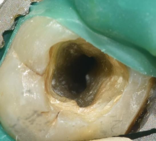
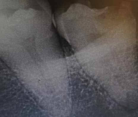
A 28-year-old female patient presented to the Department of Conservative Dentistry and Endodontics with a primary complaint of pain in the lower left posterior region. Her medical history was unremarkable. She reported suffering severe, radiating pain originating from tooth #37, induced by food impaction. The clinical examination identified a profound carious lesion on” the bucco-occlusal surface, nearing the pulp Figure 1a. The tooth had a lack of response to pulp vitality assessments and showed sensitivity upon percussion. Radiographic examination revealed a radiolucent region affecting the pulp and corresponding periapical expansion. “A diagnosis of pulp necrosis connected with symptomatic apical periodontitis was established. The radiograph indicated a solitary conical root featuring two separate canal outlines converging at the apical third, indicative of a C-shaped canal morphology Figure 1b.
Following administration of local anaesthesia and isolation with a rubber dam, an endodontic access cavity was prepared. Upon extirpation of the pulp tissue, the canal configuration was identified as Fan et al.'s C1 type. The pulp chamber was irrigated with a 3% solution of sodium hypochlorite. An electronic apex locator (E-PEX, Orikam, India) was used to calculate the working length, and radiography was used to validate it Figure 1c. Neo Endo rotary was used for biomechanical preparation instruments to a size 25 with a 6% taper (Orikam, India). Activation of irrigants was performed using an endo activator (Ultra X, Orikam, India). At the working length, a master gutta-percha cone was selected and verified radiographically Figure 1d. Later, a bioceramic sealer (Ceraseal, Meta Biomed, Korea) and the chosen gutta-percha cone were used to seal the canal. A definitive coronal restoration was subsequently applied Figure 1e.


A 54y/o female patient presented to the department with a primary symptom of pain during mastication in the lower right posterior tooth location. Her medical history was unremarkable. The intraoral examination identified a carious lesion in tooth #47, which exhibited tenderness upon percussion.
The radiographic assessment revealed a significant occlusal radiolucency in tooth #47, in close proximity to the pulp chamber. The tooth displayed a conical morphology with joined mesial and distal roots. Vitality assessment utilising cold spray and an electric pulp tester produced a delayed but affirmative reaction Figure 2b. Following these findings, the tooth was diagnosed with reversible pulpitis, and a direct pulp capping procedure was carried out. Subsequently, after three weeks, the patient presented with severe discomfort in the identical area. Consequently, root canal therapy was scheduled and explained to the patient. An access A cavity was made once adequate local anaesthesia and rubber dam isolation were achieved. It was found that the canal morphology matched the Fan et al. description of the C1 type canal architecture Figure 2a. Using an apex finder, the working length was determined, and radiographic confirmation confirmed it Figure 2c. The canal was produced to size 25 with a 6% taper using NeoEndo rotary files (Orikam, India). To create a thorough canal shape, circumferential filing using hand K-files was then carried out. Irrigation had been conducted using 3% sodium hypochlorite, activated by the EndoActivator system (Orikam, India) to improve cleaning efficacy. The master gutta-percha cone was adjusted to the working length and confirmed using a radiograph” Figure 2d. Gutta-percha and a bioceramic sealer (Ceraseal, Meta Biomed, Korea) were used in the single-cone method to seal the canal. A definitive composite repair was applied subsequent to obturation Figure 2e.


A 43-year-old woman came to the Department of Conservative Dentistry complaining of lower right posterior pain. His medical history was unremarkable. She exhibited a heightened sensitivity to thermal stimuli, particularly to heat and cold. A clinical examination identified a profound bucco-occlusal carious lesion nearing the pulp.
The tooth exhibited no response to pulp vitality testing, yet was not sensitive to percussion. The radiographic assessment indicated a radiolucent area within the pulp space, devoid of any signs of periapical disease Figure 3a. A diagnosis of pulp necrosis was established based on these findings. A C-shaped canal morphology was evident from the radiograph, which showed a single conical root with a single canal arrangement Figure 3a. An access cavity was made following the administration of local anaesthetic and the installation of a rubber dam. After necrotic pulp tissue was removed, a single circular aperture in the middle of the pulpal floor was observed; this opening matched the C4 canal pattern that Fan et al. (2004) [5] had described.
3% sodium hypochlorite was used for irrigation. Activation of irrigants was performed using an endo activator (Ultra X, Orikam, India). An electronic apex locator (E-Pex, Orikam, India) was used to determine the working length, which was then confirmed radiographically Figure 3b.
Neoendo rotary machinery (Orikam, India) was used for cleaning and shaping to a size 25 with a 6% taper. A radiograph was used to confirm that the master gutta-percha cone was adjusted to the operating length Figure 3c. The canal was sealed using a bioceramic root canal sealer (Ceraseal, Meta Biomed, Korea) in conjunction with auxiliary cones and the selected master cone.
After obturation, a definitive coronal repair was put into place Figure 3d.


Prior to obturating the root canal system using an inert substance, root canal therapy focuses on forming and sterilising the system. Insufficient canal treatment can contribute to endodontic failure [9]. A C-shaped canal denotes a distinct anatomical variant defined by a continuous groove connecting one or more canals inside teeth exhibiting joined roots. Its distinctive C-shaped cross section is what distinguishes this construction. The primary issue presented by C-shaped canals is the existence of an isthmus connecting multiple canals, complicating comprehensive debridement and full obturation [10]. Precise identification of C-shaped canals necessitates an integration of radiographic imaging and clinical assessment [11]. Chemically disinfecting the root canal system is a crucial step in eliminating microbes. Selecting the appropriate irrigation solutions is vital to achieving effective disinfection, especially in isthmus areas. For optimal chemical cleansing, we recommend activating irrigants using sonic agitation with the Endo Activator system [12]. Various materials and methods are available for obturating root canals. Finding the mixture that provides a dependable, three-dimensional hermetic seal for the canal system is crucial. In all our cases, we achieved successful clinical outcomes. The patients remained asymptomatic throughout the follow-up period, and radiographic evaluation revealed progressive signs of periapical healing. In our instance, we used bioceramic sealers selected for their increased adherence and decreased microleakage in combination with the single-cone obturation technique. These sealers are biocompatible and chemically bond to dentin due to their high calcium and phosphate content, forming a crystalline structure similar to hydroxyapatite [13].
Bioceramic sealers perform well because they conform to the complexities of the root canal anatomy, thereby providing a long-lasting seal and reducing the risk of reinfection [14]. Along with this chemical debridement and its activation, the complete removal of both organic and inorganic debris through penetration into all complex and minute dentinal structures is also necessary for successful healing of the condition. Similar results were observed in the case report by Vasileva V R et al., who used a bioceramic sealer [15]. Our clinical experiences underscore the importance of individualised treatment plans and the integration of advanced technologies to achieve the best possible outcomes. Future studies should prioritise comparative analyses of different filling techniques and materials for complex anatomies, such as C-shaped canals. Furthermore, additional investigation is needed into how specific irrigation protocols affect long-term treatment success. We think that creating a standardised procedure for the cleaning, obturation, and preparation of teeth with C-shaped canal configurations is crucial.
The case study demonstrates how to successfully treat a complicated C-shaped mandibular second molar, emphasising the value of precise diagnosis, thorough comprehension of aberrant root canal anatomy, cautious chemo-mechanical debridement, and three-dimensional obturation of the C-shaped canals to guarantee a positive long-term result.
The patients in this case series reported significant relief from symptoms following endodontic treatment for C-shaped canals. They experienced minimal discomfort during the procedure and remained asymptomatic throughout the follow-up period. Radiographic evaluations showed signs of healing, further confirming the success of the treatment. Overall, the patients were satisfied with the outcomes and the preservation of tooth function.