Oral Sphere
Journal of Dental and Health Sciences
Journal of Dental and Health Sciences
Received: 2025-03-01
Accepted: 2025-06-25
Published: 2025-07-01
Pages: 184-194
Oroantral fistula (OAF) is a pathological condition characterized by an abnormal communication between the oral cavity and the maxillary sinus. This communication leads to a persistent opening between these two anatomical regions, allowing the passage of oral fluids, food particles and microbial flora into the maxillary sinus. OAF can result from various causes including failed primary healing following dental extractions particularly in the posterior maxillary region, trauma, chronic dental infections, osteomyelitis, radiation therapy and iatrogenic complications during surgical procedures involving the maxilla. The clinical presentation of OAF often includes symptoms such as persistent or recurrent sinus infections, nasal discharge, foul taste or smell and sometimes even oro-nasal regurgitation of liquids. Diagnosis is typically confirmed through clinical examination and imaging studies such as dental radiographs or computed tomography (CT) scans, which help to visualize the fistulous tract and assess its size and extent. Management of OAF aims to close the fistulous tract and restore the integrity of the oral and sinus cavities. Treatment options vary depending on the size, location and underlying cause of the fistula. Conservative measures may include observation, nasal decongestants and antibiotics to control infection. However, surgical intervention is often necessary for definitive closure of the fistula. Surgical techniques range from simple primary closure with local flaps to more complex procedures involving bone grafts or soft tissue flaps depending on the size and complexity of the defect. This comprehensive review comprehensively explores the etiology, clinical manifestations, diagnostic modalities and management strategies pertaining to oroantral fistula (OAF).
A connection between the maxillary sinus and oral cavity occurs as Oroantral Communication (OAC) when it develops abnormally or pathologically. The failure to treat OAC results in the formation of an oroantral fistula (OAF) which produces persistent epithelium while causing serious chronic complications. An unusual pathologic condition called OAF shows an incorrect association between the oral cavity and maxillary sinus and contains epithelium as its structural lining. The development of an oroantral fistula (OAF) happens after oral epithelium migration to oroantral communication through epithelialization [1]. The formation of oroantral communication elapses during a period of 48 to 72 hours after its initial creation [2]. Communications less than 2.0 millimetres in width tend to close automatically by themselves without developing infections. Multi-surgical intervention becomes necessary when OAF defects remain larger than 5mm for more than three weeks [3].
A formed oro-antral fistula manifests an epithelial lining which incorporates both pseudostratified ciliated columnar epithelium from the maxillary antrum alongside squamous epithelium from the oral mucosa. When fistulas remain open without closure, they allow oral cavity toxins to enter thus worsening sinus inflammation and creating future complicated conditions. Medical professionals need to determine whether a sinus infection exists because proper treatment selection depends on this diagnosis together with its prevention of additional complications [4].
Treatment of maxillofacial fistula requires immediate fistula closure along with complete sinus infection assessments to achieve proper management of this condition. OAF exist in three categories based on their position within the body as alveolo-sinusal and vestibulo-sinusal and palato-sinusal lesions. The different types create connections between distinct areas between oral cavity space and maxillary sinus area [5]. Children have lower chances of oroantral communication development than adults because their maxillary sinus is smaller and underdeveloped.
The anatomical difference between children and adults helps minimize OAF risk in children [6]. This review research analyzes the clinical presentation, diagnostic methods, and treatment approaches for oroantral fistulas through an examination of published literature on this unusual pathological entity.
Etiology: OAF develops frequently after extracting maxillary molars and premolars because their roots lie near the maxillary sinus area. The extended roots of these teeth commonly penetrate inside the maxillary sinus, thereby increasing the possible risks during extraction. The procedure of extraction produces two simultaneous challenges by causing damage to the sinus tissue and creating accidental exchange routes between oral cavity and maxillary sinus spaces.
The establishment of an OAF becomes possible after this breach because the pathway from the oral cavity to the maxillary sinus persists, which may develop into chronic sinus conditions without appropriate management. Root canal procedures of specified teeth also risk OAF formation because they produce the same type of mechanical damage to sinus lining tissue. Maxillary antrum bone measures between 12 mm and no bone at all in thickness when surrounding the roots of maxillary molars and premolars. The insufficient bone tissue creates elevated OAF risks especially when extractions or root canal procedures performed [4].
The development of OAF can occur because of maxillary tuberosity fractures and periapical infections along with pathological conditions such as cysts and tumors. Iatrogenic injuries together with Paget's disease act as two additional conditions responsible for creating OAF [7]. OAF development can be initiated by maxillofacial trauma along with osteomyelitis and radiation. Posterior maxillary dental implant procedures sometimes cause OAC as a surgical complication or post operative complications. Indirect sinus lift before dental implants in this area has sometimes led to the development of OAF [8].
Signs and Symptoms: Signs and symptoms can emerge in either acute or chronic presentations. Patients with OAF commonly experience sudden symptoms which include epistaxis, pain and fluid or air leakage from the fistula and voice alterations. The symptoms of chronic OAF appear as persistent pain combined with uncontrolled fluid leakage and presence of antral polyps along with postnasal drip and dysgeusia and changes in voice quality along with ear pain and mucopurulent nasal discharge [9].
Nose-blowing test - The patient should close their nostrils with their hand and keep their mouth open during forced nasal expiration to test for OAF using the nose-blowing method. This procedure reveals OAF when the doctor hears audible whistling, accompanied by bubbles, or visualizes fluid around the hole.
Cotton-wisp test/Butterfly test - A cotton wisp or butterfly test enables the operator to verify potential oroantral fistula by placing it close to the orifice. Detection of OAF relies on negative pressure that can draw the cotton wisp into the sinus opening.
Mouth mirror test - To check for a possible OAF, the mouth mirror is placed near the orifice. Air escaping from a fistula can produce fogging on a mouth mirror which confirms the existence of OAF. When air flows from the oral cavity to the maxillary sinus, it causes mirror condensation on the surface [10],[11].
A panoramic view helps detect alveolar defects, and the Waters view provides better detection of maxillary sinus infections.
CT scan examinations, as well as cone-beam computed tomography (CBCT), can be diagnostic tools for OAF. CBCT as a modern radiographic tool and is particularly effective in diagnosing and detecting oroantral defects with high precision. Additionally, these imaging techniques enable the identification of sinus mucosa thickening or opacification, assessment of nasal meatus aeration, and evaluation of ethmoidal air cells and other sinuses for pathological conditions [12].
Closure of OAF can be determined by various factors that impact both the surgical outcome and post-closure rehabilitation. These include seniority and defect size. While smaller OAFs (less than 2mm in diameter) may spontaneously close, larger defects (> 4mm) tend to persist and necessitate surgical closure [13].
For Oroantral Fistula (OAF) closure to be successful, it is critical to follow some basic guidelines to achieve proper healing without recurrence. The first principle addresses the issue of ensuring that the sinus is infection-free. An infection in the maxillary sinus can severely obstruct healing, as the bacteria and inflammation present tissue regeneration complications and further issues. Therefore, thorough evaluations of the sinus must be completed prior to surgery. These evaluations must demonstrate the infection is absent, and if needed, proper Medical Management (antibiotics or decongestants) is used to treat infection or sinus pathology [13],[14].
The second OAF closure principle relates how to perform closure `tension freesly'. This is done by obtaining a broad base, well vascularised soft tissue flap. Closing defects with well vascularised tissue is neccessary to provide adequate blood flow to healing tissue which is important in tissue regeneration and flap healing which prevents necrotic complications. The flap should completely cover the intact bone and the wound edge should be closed in a way that does not stretch or pull on the adjacent tissues. Tension-free closure facilitates healing, prevents complications, and scarring [14].
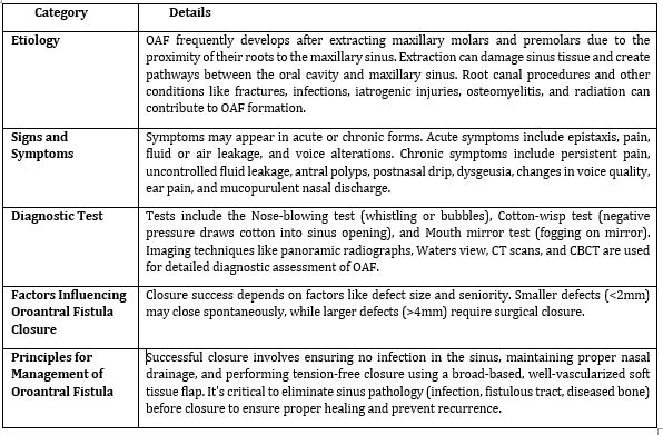
The specific aspect or topic related to Oroantral Fistula (OAF) being discussed, including its etiology, symptoms, diagnostic tests, influencing factors for closure, and management principles is discussed in Table 1.
| Category | Details |
|---|---|
| Etiology | OAF frequently develops after extracting maxillary molars and premolars due to the proximity of their roots to the maxillary sinus. Extraction can damage sinus tissue and create pathways between the oral cavity and maxillary sinus. Root canal procedures and other conditions like fractures, infections, iatrogenic injuries, osteomyelitis, and radiation can contribute to OAF formation. |
| Signs and Symptoms | Symptoms may appear in acute or chronic forms. Acute symptoms include epistaxis, pain, fluid or air leakage, and voice alterations. Chronic symptoms include persistent pain, uncontrolled fluid leakage, antral polyps, postnasal drip, dysgeusia, changes in voice quality, ear pain, and mucopurulent nasal discharge. |
| Diagnostic Test | Tests include the Nose-blowing test (whistling or bubbles), Cotton-wisp test (negative pressure draws cotton into sinus opening), and Mouth mirror test (fogging on mirror). Imaging techniques like panoramic radiographs, Waters view, CT scans, and CBCT are used for detailed diagnostic assessment of OAF. |
| Factors Influencing Oroantral Fistula Closure | Closure success depends on factors like defect size and seniority. Smaller defects (<2mm) may close spontaneously, while larger defects (>4mm) require surgical closure. |
| Principles for Management of Oroantral Fistula | Successful closure involves ensuring no infection in the sinus, maintaining proper nasal drainage, and performing tension-free closure using a broad-based, well-vascularized soft tissue flap. It's critical to eliminate sinus pathology (infection, fistulous tract, diseased bone) before closure to ensure proper healing and prevent recurrence. |
Introduction of buccal flap by Axhausen in 1930 and serve as the reliable technique for closing small to moderate fistulas. Coverage of fistula involves a vertical incision along the buccal region with a thin layer of the buccinator. While relatively straightforward to execute, careful handling is necessary due to the thin nature of these flaps. Notably, one advantage of this method is its applicability in patients with significant alveolar resorption. Various types involve advancement, rotated, transversal and sliding flaps [15].
Rehrman introduced the Buccal Advanced Flap technique in 1936, making it one of the oldest and most frequently used methods for treating OAF [16]. Surgeons often opt for this method as the primary treatment for closing small communications or minor fistulas that require simple suturing.
This technique involves excising the epithelialized margins of the oroantral fistula and creating two diverging vertical incisions that extend from the extraction site to the buccal vestibule. A broad-based trapezoidal mucoperiosteal flap is then elevated, positioned over the defect and sutured from the buccal to the palatal mucosa using horizontal mattress sutures. Advantages include high survival rate and sufficient blood supply [17]. However, a notable disadvantage is the potential reduction in buccal sulcus depth post-surgery, which can decrease retention and cause discomfort for denture wearers [16]. Improper closure of large defects are due to weak perfusion of the flap [18].
Moczair introduced the Buccal Sliding Flap technique as an alternative method for the closure of alveolar fistulas. One advantage of this approach is its minimal impact on depth of buccal sulcus achieved by shifting the flap one tooth distally. Significant dentoalveolar detachment required for the shift may lead to gingival recession and periodontal disease. This is suitable for edentulous patients . Rehrman and Moczair flaps lead to post-operative swelling as a consequence of mucoperiosteal flap reflection [13]-[18].
The Buccal Fat Pad (BFP) was introduced by Egyedi as a pedicled graft technique for the closure of OAF, offering a reliable and effective method due to its rich vascular supply and ease of mobilization.
This technique gained popularity following Tideman's study, demonstrated that BFP epithelializes within 3-4 weeks. The procedure resembles that of the buccal flap technique, beginning with a circular incision around the fistula.
Subsequently, two vestibular incisions are made in maxillary tuberosity and then blunt dissection done beneath the periosteum. In this technique, the fat pad is gently mobilized and transferred to the recipient site, where it is sutured to the palatal tissue. The fat pad is typically left uncovered in the oral cavity, allowing it to epithelialize naturally over time [19]. This flap offers several advantages including adequate vascularity, a high success rate, a lower risk of infection, rapid epithelialization of the exposed fat, minimal donor site morbidity and versatile application. These benefits make it a preferred choice for closing medium-sized maxillary defects effectively [20].
However, risk of recurrent fistula and graft necrosis when it is used for large defects. A history of radiation therapy should be carefully evaluated, as previous radiation can affect the size and mobility of the BFP. Additionally, concerns about the safety of BFP technique arise due to the potential risk of injuring the pterygomaxillary space during the procedure [21].
Palatal flaps used for the closure of oroantral fistulas include various types such as rotation-advancement flap, hinged flap, pedicle island flap, anteriorly based flap, straight-advancement flap, submucosal soft tissue pedicle flap and submucosal island flap [22].
The straight-advancement flap is typically used for closing minor palatal or alveolar defects, as it doesn't offer significant lateral coverage mobility. The pedicle island flap was introduced by Henderson for closure of OAF in 1974. This flap offers advantages such as excellent blood supply, bulk and mobility. Dental surgeons choose the palatal island flap as their preferred method for treating oroantral fistulas because it provides a simple and versatile approach with effective mobility that makes it workable for different sized defects [13],[22].
Hara and Ito developed the submucosal connective tissue pedicle flap, which divides tissue into two parts by separating the mucosal layer above and the connective tissue layer beneath. This surgical method was developed to address the issue of donor site bone exposure, as it provides an enhanced method for wound closure protection. The complete thickness of the palatal flap gets separated into two different sections consisting of mucosa on top and connective tissue underneath. The fistula closure begins by deploying the connective tissue layer and placing the mucosa back to its native site to achieve primary suture of the tissue. All study reports indicate that donor site healing is complete within one month. The complexity of dissection, combined with potential complications related to blood supply, makes this procedure difficult to perform [23].
Dergin et al (2016) [17] established this method for treating oral antral fistula occurring between the second and third molar area. The elastic and easy-to-manipulate flap demonstrates excellent adaptability while stopping the formation of dog-ears and flap folding. The procedure protects donor site bone tissue through the separation of the flap into two layers that consist of mucosa along with underlying connective tissue which aids tissue preservation and healing.
The first step involves removing the fistulous wall and clearing granulation tissue using a curette. An H-shaped, window-like incision is then made in the palatal mucosa, approximately 4 mm away from the gingival margin to access and prepare the site for closure. Arterial connective tissue flap is then dissected and positioned through palatal tunnel maneuver. The surgeon performs tension-free sutures for the flap area. Early wound healing, along with reduced discomfort, occurs because the epithelial layer can be returned to the donor site with this procedure. The healing process of the donor site typically takes about one month. The procedure has several disadvantages, including the potential for damage to the blood supply, complex dissection requirements, and a dependence on a skilled surgeon for successful execution [24].
The Palatal Rotational Flap with complete thickness represents a common treatment approach for OAF which exceeds 10 mm or needs delayed repair. PRF represents a different approach because it requires the removal of total tissue layers from both the mucosa and periosteum. The preservation of buccal vestibular depth makes this technique the better choice than buccal flap procedures [25].
Hooking the design of the flap around the greater palatine artery ensures proper blood supply. The surgeon performs a circular removal of the fistulous tract when needed. The surgical procedure involves developing a wide flap extending from the full palatal thickness that incorporates the greater palatine artery which is then carefully rotated to enclose the defect. The process of suturing requires either a collagen sponge or a palatal splint to help secondary epithelialization while promoting healing of the donor site. Rotational movement of the flap becomes limited because of thick keratinized tissue in situations where the fistula exists in the maxillary tuberosity region. The procedure offers several benefits due to its substantial mass and solid keratin formation, accompanied by a sufficient blood supply and valuable conservation of vestibule depth [26].
The surgical technique is restricted by flap movement during procedures involving thick keratinized tissue, which becomes more pronounced when fistulas exist in the maxillary tuberosity region [27].
This procedure has two main disadvantages: the greater palatine artery restricts flap movement, and a dog-ear may occur at the pivot point during rotation, potentially damaging the blood supply and affecting healing results. Excision of the formation of a dog-ear is necessary for better adaptation. Additionally, re-epithelialization may be needed due to bone exposure on the hard palate. Risk of necrosis at the donor site is observed in medically compromised patients.
V-shaped excision by Kruger at the flap's greatest bend to prevent wrinkling and folding. Epithelialization of the exposed palatal bone typically occurs within two weeks [28]. It is advisable to employ a palatal stent after surgery to stabilize the flap and mitigate edema [28].
The palatal hinged flap is an effective technique for closing small fistulas of the hard palate, typically those less than 2 cm in diameter and is usually performed as a single-stage procedure. The procedure starts by elevating a complete flap immediately adjacent to the fistula and aligning it along one edge. The flap is then rotated like a hinge over the fistula, with its buccal surface positioned on top within the defect. An advantage of this technique is that it leaves only a small raw area for granulation tissue to form, ensuring efficient closure of the OAF [29],[30].
The palatal pedicled island flap is another single-stage local flap, valued for its rich vascular supply, substantial bulk and excellent mobility. The flap is elevated by creating incisions specifically tailored to provide the required volume of donor tissue for effective fistula resurfacing. The palatal pedicled island flap incisions typically placed between the hard and soft palate junction or less than 5 mm from the teeth to enable proper tissue movement and protect blood supply.
The strategic area facilitates the removal of tissue while maintaining complete functionality of the vascular pedicle and provides 180° mobility of the harvested flap. Numerous surgeons choose the palatal pedicled island flap procedure due to its multiple advantages, including ease of use, versatile design, and movement capabilities [31]. Posterior fistulas benefit best from palatal pedicled island flap treatment because this surgical approach supplies a broad section of well-vascularized tissue. The hard palate location for the donor site enables quick healing along with minimal tissue damage to the surrounding area. The use of this surgical approach becomes unfavourable when addressing anterior defects, as advancing the flap puts stress on the vascular pedicle, which could damage blood flow [32].
The palatal anteriorly based flap stands as a vital method to repair both large oroantral fistula and tuberosity region defects. The surgical procedure requires shifting mucoperiosteum laterally from the posterior area of the hard palate region. A properly raised flap successfully covers extensive defects by closing the entire area and protects raw tissue surfaces, thereby enhancing the body's healing process [33].
The tongue functions perfectly as a donor site because it possesses both flexibility and an appropriate blood supply, along with suitable placement. Relocating tongue flaps occurs based on the specific defect location either from the dorsal side or from the ventral or lateral aspects [25]. Lateral tongue flap achieves effective results for large oroantral fistula closure through treatment success levels between 85% and 95.5% [26]. The surgical procedure of tongue flap surgery leads to wound dehiscence along with hematoma formation followed by shortterm loss of sensation and taste. It requires both general anesthesia and additional surgeries for successful outcomes [34].
The temporalis muscle flap functions as a distant flap to effectively address defects in the orofacial region while specifically aiding single-stage treatment of large oroantral communication. The surgical procedure requires precise cutting of temporalis fascia above the zygomatic arch which enables the flap to rotate. Surgeons guide the flap through a surgically made passage in the infratemporal fossa for successful closing of the oroantral fistula. The temporalis muscle flap demonstrates superior performance compared to conventional alternatives, as it produces minimal bulk while offering enhanced vascular function, improved flexibility, and preventing both functional and aesthetic defects. The close location of this flap to the oral cavity enhances its effectiveness according to research [35].
The treatment of OAF typically involves bone autografts for cases with defects exceeding 10 mm in size or when conservative procedures fail to achieve proper closure [29],[30]. Autografts can be sourced from various sites including the extracted socket, chin, retromolar area, zygomatic process or distant location such as the iliac crest, to effectively repair maxillary bony defects. Harvesting bone from intraoral donor sites offers the added advantage of reducing postoperative discomfort and the demands on patients' recovery [36].
This procedure for OAF closure may involve the removal of third molars, which could potentially affect patient acceptance [31]. A drawback of intraoral bone autografts is the limited availability of bone compared to chin bone grafts. This procedure involves making an incision medial to the external oblique ridge, followed by the elevation of a mucoperiosteal flap. During the procedure, surgeons use a micro-reciprocating saw to create osteotomies by lifting carefully to avoid nerve entrapment. Post-trimming of osseous irregularities the surgeon moves the flap into position while finishing the repair through tissue suturing [37].
The zygomatic bone serves as an ideal bone donor site for OAF closure when surgeons need to collect small volumes of bone tissue. An incision is made above the mucogingival junction, enabling surgeons to lift a full-thickness flap before removing bone material from the zygomatic rim area. This procedure offers key benefits, as it reduces surgical duration while allowing for a shorter distance to the deficient site and providing diminished patient discomfort [38].
The use of auricular cartilage is continuing to evolve as a treatment approach for OAF. The surgical procedure begins by raising a flap through the defective tissue, followed by a semicircular cut through the conchal cartilage to collect the graft material. The harvested auricular cartilage is then shaped to fit and sutured onto the defect site. Subsequently, the mucoperiosteal flap is advanced and sutured in conjunction with the palatal tissue to ensure secure closure. This technique offers biocompatibility, high resistance to infection and easy harvest without requiring vascularization for integration, thus reducing graft failure rates. However, a potential drawback is the formation of defects at the donor site [39].
Septal cartilage has been documented as particularly effective for larger oroantral fistula [48]. This technique begins by raising a buccal mucoperichondrial flap, typically extending from two teeth mesial to one tooth distal of the fistula. An incision is made at the caudal end of the septal cartilage, followed by elevating a mucoperichondrial flap on one side. A cartilage island is then outlined, carefully dissected free, trimmed and inserted into the defect as a horizontal plate. Saleh et al reported a success rate of 95.7% with the use of septal cartilage for OAF closure [40].
OAF has been used with allogenous materials such as lyophilized fibrin glue and dura . Fibrin glue is lyophilized with a collagen sheet and injected into the socket. It is used as a straightforward technique of closing oroantral fistula. Its use, however, has its risks such as transmission. It is associated with viral hepatitis and requires additional preparation time before application [41]. Dura is rehydrated in saline in order for it to regain pliability and is placed over the bony margins of the defect. It is sutured and wrapped in a plastic plate for protection. Although the technique is simple, this carries the risk of transmitting pathogens which is notable.
Xenograft material based on lyophilized porcine dermis has established itself as a promising option for OAF closure. The graft procedure can be performed either by exposing the material directly to oral contact or by applying it beneath protective flaps. These grafts exhibit both wide application range and favourable therapy results [38],[39]. Researchers argue that flap coverage may not always be necessary for oroantral fistula closure. Collagen offers the advantage of being naturally incorporated into granulation tissue, which eliminates the need for subsequent removal, simplifying the healing process [42]. Few researchers also successfully achieved closure of hard and soft tissue defects using Bio-Gide® and Bio-Oss® without requiring donor site surgery. However, a notable drawback of this technique is the necessity of creating a flap to cover the "sandwich" graft, which adds complexity to the procedure [40]-[42].
Kitagawa et al. (2003) [41] advocated for the transplantation of third molars as an effective method for OAF closure. They reported successful outcomes in two cases involving the immediate transplantation of upper and lower third molars to seal the defect . Firm pressure and gentle tapping ensured tooth stabilization, resulting in complete closure. However, this approach has drawback including the necessity for a well-developed third molar with a suitable shape and size for transplantation. Additionally, there is a risk of complications, such as ankylosis and root resorption, if the procedure is not executed with precision [40],[41].
Interseptal alveolotomy involves removing the interseptal bone and fracturing the buccal cortex toward the palate, followed by suturing to achieve soft tissue closure. This method promotes spontaneous postoperative healing, reduces swelling and ensures tension-free closure. However, limitations include the requirement for a space of less than 1 cm between adjacent teeth and an adequate alveolar ridge. Additionally, incomplete fractures can lead to inflammation and imperfect closure, posing risks to the healing process [22], [42].
Waldrop and Semba introduced the guided tissue regeneration for OAF closure [11]. This method entails placing an absorbable gelatin membrane over OAF, followed by the application of allogenic bone graft material. A nonresorbable ePTFE membrane is then used to cover the graft. The ePTFE membrane is removed after eight weeks, with closure confirmed clinically through bone formation. Despite its effectiveness, this technique has drawbacks, including the need for an additional surgical procedure to remove the ePTFE membrane and the requirement for a full-thickness flap, which adds complexity and patient discomfort [42].
Selecting the optimal surgical approach for OAF closure involves careful consideration of factors such as fistula size, location, adjacent teeth, alveolar ridge height and patient health. Smaller fistula (5 mm), advanced options include per-oral buccal fat pad flap, pedicled buccal fat pad, modified submucosal connective tissue flap, distant flap, autogenous bone graft, allogenic material, synthetic substitutes or the Bio-Gide®-BioOss® sandwich technique. The choice of procedure should align with the indication and the surgeon's expertise. For less experienced practitioners, simpler techniques such as PTFE membrane or Rehrmann flap may be preferable to more complex options, such as bone graft transplantation. Dentists must thoroughly assess their skills and select the most appropriate method to ensure successful closure of the OAF.