Oral Sphere
Journal of Dental and Health Sciences
Journal of Dental and Health Sciences
Received: 2025-04-01
Accepted: 2025-03-13
Published: 2025-04-01
Pages: 80-94
Introduction: Nearly 3.5 million people worldwide suffer from dental caries. Untreated dental caries has been identified as a prevalent oral health condition that raises the burden on oral health and may cause other systemic illnesses. Smartphones and Artificial Intelligence are now widely used in many healthcare domains, including diagnosis and treatment planning. So, this umbrella review aims to appraise the existing evidence on the effectiveness of various algorithms of Artificial Intelligence in diagnosing dental caries from previously published systematic reviews.
Material and Materials: A broad electronic search was done among various databases that focus on existing systematic reviews on dental caries detection or diagnosis using various algorithms of Artificial Intelligence. The quality assessment of the included systematic reviews was done using the AMSTAR-2 checklist and the risk of bias was done using the ROBIS tool.
Results: Among 503 reviews, 9 were included in this umbrella review. Based on the AMSTAR-2 checklist, 7 reviews were stated as high quality and 2 reviews as moderate quality; and the all the reviews were stated to have low risk of bias. The sensitivity, and specificity values of machine learning models employed in diagnosing dental caries ranged from 71% to 99% and 90% to 94% respectively, and accuracy ranged from 77.4% to 90%, whereas the sensitivity, specificity values for deep learning models ranged from 78.6% to 87% and 78.4%. to 87.8% respectively.
Conclusion: The diagnostic performance of artificial intelligence models varies between machine learning and deep learning algorithms. Based on these findings, machine learning has shown promising results in diagnosing dental caries.
Oral diseases are a significant global health burden affecting individuals throughout life. But when such conditions are compiled to ignore or delay treatment, the treatment creates discomfort or pain, and invasive operations that need to be a payment that increases the financial burden. Most conditions, however, can be caught early enough and treated to prevent or reverse the disease. Approximately 3.5 billion people have oral disease, 2 billion people have caries of the permanent teeth, and nearly 514 million children—0.7% of the world’s population—are affected by caries of the primary teeth [1].
In the Global Burden of Disease 2019 report, untreated dental caries in permanent dentition was the leading oral health condition reported.
According to the WHO Global Oral Health Status Report 2022, oral disease is estimated to affect approximately 3.58 billion people worldwide. Around 2.5 billion individuals show signs of caries in permanent teeth, and nearly 514 million children are affected by caries of the primary teeth [2].
The overall prevalence rate for dental caries in India is 54.16%. The specific prevalence was 62% in subjects above 18 and 52% among the age group of 3–18. The approximate prevalence of malocclusion in the mixed dentition is 58%. By region, it was highest in Western India, affecting 72% of the population [3].
Recent interest has, therefore, shifted towards the early detection of carious lesions to counter complex treatment procedures. Dental caries can be diagnosed using a wide range of methods. Although the visual-tactile method is widely accepted as the first-line diagnosis, it has not enabled the identification of various lesions with interproximal surfaces. The Radiographic Approach for Caries Detection is used to recognize this limitation of the visual method.
Numerous radiographic techniques can be used for caries detection, but posterior bitewing, periapical, and panoramic views are the most commonly used. “In any case, the entire maxillary and mandibular arches can be visualized by panoramic imaging, but in panoramic imaging, the incipient carious lesions are poorly defined” [4].
Various diagnostic criteria and techniques for early detection of dental caries are available. However, most people are still hesitant.
Red is the first phase, either not visiting the dentist or avoiding setting the stage, and the scrutiny in the next phase will cost invasive treatment or the loss of a tooth altogether. Furthermore, put in the context of where we are today, AI is an alternative that might enhance our ability to detect oral diseases in their onset stages [5].
Some AI applications in healthcare can further improve the accuracy of cancer diagnosis, early diagnosis of fatal blood disorders, virtual health assistants, automation of redundant healthcare tasks, management of medical records, robot-assisted surgery, automated image diagnosis, and improved healthcare access.
AI systems need to undergo training on similar sets of samples (for instance, subjects), relationships between the features of the subjects, and the relationships between the samples and the outcome of interest before they can be integrated within healthcare applications (this training can be undertaken through datasets that represent clinical activities, e.g., screening, diagnosis, treatment assignment, etc.) [6].
AI in diagnostics and treatment planning are the other fields of dentistry where AI has entered. For example, the integration of AI in analyzing the disease has been reported in the various advances in radiographic techniques for diagnosing dental caries. Photographs made by machine learning can also be used to diagnose dental caries.
This has significantly increased in recent clinical studies and reviews investigating the performance of Artificial Intelligence algorithms in diagnosing dental caries. So, we wondered if we could summarise our data and whether an umbrella review could indicate the applicability of various AI algorithms (machine learning deep learning) to diagnosing dental caries.
The objective of this umbrella review is to review the available evidence for systematic reviews assessing the diagnostic effectiveness of AI (ML and DL) for identifying carious lesions.
This study protocol was based on Preferred Reporting Items for Overviews of Reviews (PRIOR) guidelines.
An umbrella review was conducted based on the PICO statement
Participants (P): Existing literature evidence of Artificial Intelligence in diagnosing dental caries among intraoral images and radiographs.
Exposure(E): AI to detect dental caries
Comparison (C): Not applicable
Outcome (O): Effectiveness of AI in caries detection.
It includes systematic reviews that assessed the intraoral clinical images of patients, as well as periapical, near-infrared light trans illumination, and bitewing radiographical images, focusing on dental caries in patients of all ages and without any other disorders.
Systematic reviews that assessed the ability of various Artificial Intelligence algorithms to detect, diagnose, and predict dental caries based on images and radiographs were included.
This review included systematic reviews that assessed AI algorithms for detecting dental caries using clinical images and radiographs.
Systematic reviews and meta-analysis.
Reviews that investigated the diagnostic ability of dental caries by Artificial Intelligence.
Reviews only in the English language were included.
Reviews from January 2019 to June 2024 were included.
Systematic reviews investigating the scope of Artificial Intelligence, various techniques of AI used for treatment planning, and diagnosing purposes for oral diseases other than dental caries. AI used in various fields of healthcare other than dentistry was excluded.
Narrative reviews, scoping reviews, and primary studies, such as observational, analytical, cross-sectional, retrospective, prospective cohort studies, randomized controlled trials, letters to the editor, and clinical guidelines related to applications of AI in dentistry, were excluded.
Relevant studies were identified through a broad electronic search in PubMed, Scopus, Web of Science, EMBASE, Google Scholar, Cochrane database of systematic reviews, and Trip databases. Studies published between 1998 and June 2024 were included, and the search for relevant studies was performed up to June 30, 2024. Corresponding authors of these potentially pertinent articles without full text were contacted via e-mail and requested to send us a copy of the article. A search string was used based on the statement.
Search terms were Artificial Intelligence, dental caries, machine learning, and oral photographs (MeSH terms). The same applies to the Boolean operators and OR. “Systematic review” filters were also used. The PubMed searches were: Artificial Intelligence AND dental caries, Artificial Intelligence AND diagnosis, Dental caries AND diagnosis AND oral photographs, and oral photographs AND Artificial Intelligence with the filter “only systematic reviews.” The exact keywords were searched in the Cochrane database with no filter. In Google Scholar, the keywords were Artificial Intelligence, dental caries, diagnosis, and more advanced search with the features (With all of the words: Artificial Intelligence, dental caries, diagnosis); (With the exact phrase: Artificial Intelligence, dental caries, diagnosis); (With at least one of the words: dental caries) were used. The only filters retained were “review articles.” Table 1 presents a detailed search strategy utilizing the keywords across varied databases.
The initial search resulted in 370 reviews based on the title searched in five databases: PubMed, Cochrane Database of Systematic Reviews, Scopus, Web of Science, and Trip database. Out of which PubMed produced 366 reviews, the Cochrane Systematic Review database yielded one review, and the Trip database gave three reviews. The additional hand searching includes the search of grey literature, and Google Scholar gave 133 reviews. Among these results, 376 reviews were eliminated due to duplication, and 358 were eliminated after reading the abstract and title, which were irrelevant. In this, 19 systematic reviews were assessed for full-text reading, and 10 reviews were excluded based on specific criteria like the records that were not relevant to AI in diagnosing dental caries (N = 2), records discussing the potential of AI in treatment planning (N =3), non-availability of full text (N = 1) and those of study designs including interventional, observational, RCT, cross-sectional, meta-analysis (N = 4). Based on the inclusion and exclusion criteria mentioned, nine systematic reviews were selected for the review. The selection process was primarily done by two investigators independently.
The first author screened each review for eligibility by the title and abstract, followed by full-text screening. At the same time, the second author screened those articles and found good agreement among them. This umbrella review includes systematic reviews without meta-analysis, which assess the diagnostic ability of artificial intelligence algorithms in detecting dental caries using intraoral clinical images.
All systematic reviews from 1998 to 2024 published in English were included. This was accomplished by having each investigator independently review the study’s title and abstract. After the article/ review search using the keywords and Boolean operators, the individual studies from each database mentioned above were noted separately. Subsequently, references to those studies and duplicates were managed using the Zotero software. The software mentions duplicate studies and can be easily identified and merged into a single study/ review. The review covered studies that satisfied the minimum eligibility requirements. After the full-text publications were screened for eligibility, reviews and duplicates that didn’t pertain to the study topic were removed. The first two authors analyzed the studies, and the consensus from the third author resolved the discrepancies between the first two authors. A detailed description of the eligible studies is described in the PRISMA flowchart Figure 1.
Methodological quality was rated for systematic reviews using the AMSTAR-2 checklist, a 16-item tool containing seven critical and nine non-critical items. AMSTAR-2 then categorizes reviews into high (zero or one non-critical weakness), moderate (more than one non-critical weakness), low (one critical defect with or without non-critical weaknesses), or critically low (more than one essential weakness).
Seven systematic reviews scored high quality, two were of moderate quality, and none scored poor or critically low. The final analysis included nine systematic reviews. Data was independently collected and compared by two reviewers.
The data of interest included authors, year of publication, objectives; databases searched, included studies, risk of bias assessment tools, summarised results, and comments.

| Database | Search Terms | |||
|---|---|---|---|---|
| PubMed | #1 AND #2 AND #3 | AND #4 OR #5 | ||
| #4: ((systematic review [Title/Abstract]) OR (meta-analysis [Title/Abstract])) | ||||
| #2: (((dental caries* [Title/Abstract]) OR (caries [Title/Abstract]) OR (tooth decay [All Fields])) OR (AI [Title/Abstract]) OR (dental caries [MeSH Terms]) OR (decayed teeth)) | ||||
| Scopus | #3: Artificial Intelligence AND machine learning AND deep learning AND dental caries OR caries detection AND reviews | |||
| #1: Artificial Intelligence AND deep learning AND dental caries OR caries detection AND reviews | ||||
| #1: Artificial Intelligence AND deep learning AND dental caries AND caries detection AND intraoral images AND review OR systematic review AND meta-analyses | ||||
| Google Scholar | #3: Artificial Intelligence OR AI algorithms AND dental caries AND diagnosis AND intraoral images AND reviews | |||
| #1: Artificial Intelligence OR neural networks AND AI algorithms AND dental caries AND intraoral images AND caries detection AND reviews |
The umbrella review highlighted the potency of Artificial Intelligence diagnostic ability/ efficacy with oral photographs in diagnosing dental caries, as highlighted by the systematic reviews included in the umbrella review. A total of 370 records were identified via a database search and 113 by other sources. Nine articles were included in the 19 articles evaluated for inclusion in the review. The included systematic reviews are described in detail in the PRISMA flowchart Figure 1.
The inclusion of systematic review characteristics is summarized in Table 2 which outlines the years from 2015 to 2024. Cross-sectional, cohort (prospective and retrospective), in-vivo studies, case-control, case studies, case reports, and clinical and experimental trials were most of the types of studies included in the systematic reviews, and only one randomized controlled trial was included in a review by Khanagar et al. This review included only the articles in the English language Table 1,2.
The median output stated that the AI models tested showed caries diagnosis accuracies between 76% and 88.3%, sensitivity between 73% and 90%, and specificity between 61.5% and 93% [8]. The studies’ caries prediction accuracies ranged from 83.6% to 97.1%.
Another review [11] stated that caries lesions of any depth compared to sound teeth were detected, with the F1 Scores ranging from 68.3% to 94.3% for the classification task. For the detection task, the F1 scores varied between 42.8% and 95.4%. For caries detection on images captured with professional cameras, the F1 Score was between 68.3% and 95.4%, 78.8% to 87.6% for intraoral cameras, and 42.8% to 80% for smartphone pictures.
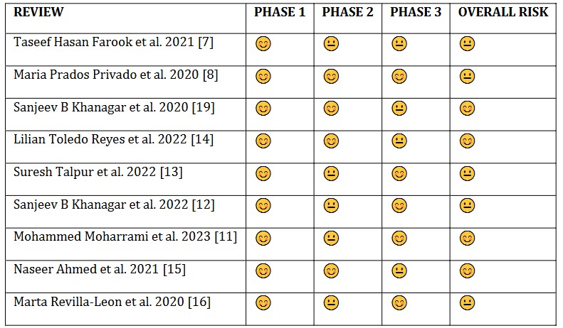
The methodological quality assessment of the systematic review included in our umbrella review, which was assessed using the AMSTAR-2 checklist, is shown in Table 3. Among the included systematic reviews, two reviews [17],[10] had “moderate quality,” whereas the remaining seven reviews [9],[11],[16],[15],[14],[13],[12] were reported as “high quality.”
The risk of bias assessment of the included studies using the ROBIS tool is shown in Table 3,4. Phase 2 and Phase 3 scoring was done in our review for all nine included systematic reviews. Based on the signaling questions of each domain, the overall risk in the included studies was calculated. One review [17] showed an “unclear risk of bias” in synthesizing the finding domain of Phase 2. After summarising the responses of each domain in both phases, we concluded that all the included reviews [9]-[16] were found to have a “low risk of bias.
| Author/year | Aim/objective / Research question | Study Design | Included Primary Studies (in sr) | Risk of bias tools used for quality assessment | Meta-analysis done / not | Outcome | Summary of results |
|---|---|---|---|---|---|---|---|
| Maria Prados-Privad et al. (2020) | What neural networks are used? (1) How is the database used to construct those networks? (3) How are lesions detected, in which teeth are they detected? (4) What are the neural networks' outcome metrics and values? | Systematic review | Cross-sectional | Cochrane Handbook Tool | No | Seven studies explained the diagnostic performance of artificial intelligence models to detect and diagnose dental caries. | The diagnostic performance varies depending on the algorithms used, with sensitivity of 89%, precision from 0.615 to 0.99, and AUC from 0.74 to 0.971 (mean ± SD 0.815 ± 0.11). Verification of generalizability and reliability is necessary. |
| Taseef Hasan Farook et al. (2020) | To evaluate the clinical effectiveness of AI models for diagnosing dental diseases based on radiographs. | Systematic review | Cross-sectional, Cohort, Case study | Joanna Briggs Institute Critical Appraisal for Diagnostic Test Accuracy (JBI-DTA) checklist and GRADE | No | AI models based on radiographs demonstrated variable diagnostic accuracy for different dental diseases. | AUC ranged from 0.74 to 0.93 for various radiographs (proximal and periapical), with sensitivity and specificity variations. AI serves as an “add-on” to existing diagnostic tools. |
| Sanjeev B. Khanagar et al. (2021) | To identify the development of AI applications in dentistry and evaluate their performance in diagnosis, clinical decision-making, and predicting the prognosis of treatment. | Systematic review | Cohort – prospective and retrospective, Cross-sectional, RCT | QUADAS-2 | No | A study showed that AI models, including gene polymorphism-based models, predicted dental caries with high accuracy. | Sensitivity of 90%, specificity of 96%, and overall accuracy of 93% (p < 0.0001), with an AUC of 0.970. Prediction accuracy ranged from 90.9–98.4%, and an ML model for early childhood caries had an AUROC of 0.774–0.785 |
| Author/year | Aim / Objective / Research Question | Study Design | Included Primary Studies (IN SR) | Risk oF Bias tools used for quality assessment | Meta-analysis Done / Not | Outcome | Summary of results |
|---|---|---|---|---|---|---|---|
| Naseer Ahmed (2021) | To understand the current trends of AI in dentistry and its application, a systematic review was carried out on studies that have discussed different modalities of artificial intelligence. | Systematic review | Clinical trial, experimental, cohort, case-control | Newcastle-Quality assessment scale (NOS) | No | The artificial neural network (ANN) models using biting show 97.1% accuracy for dental caries photos, 94.5% sensitivity, and 85-90% accuracy. | AI techniques assist dental practitioners in numerous ways, from decreasing chair time to saving time during surgery. AI can be successfully used for patient diagnosis, clinical decision-making, and predicting oral diseases. |
| Lilian Toledo Reyes et al. (2022) | To systematically evaluate the performance of AI algorithms and its prognostic prediction of dental caries and machine learning, and how these help improve both. | Cross-sectional | - | QUADAS-2 | No | The AUC values range between 0.745 to 0.987. Sensitivity scores were higher in tests where the system provides a better prediction. | The use of these technologies for the diagnosis and prognosis of dental caries helps improve patient outcomes when integrated into clinical dentistry. |
| Suresh Talpur et al. (2021) | To conduct a review of studies concerning artificial intelligence models for diagnosing dental caries and machine learning. | Systematic review | In vivo | - | No | Based on various algorithms, the neural network algorithm showed accuracy above 90% for detecting dental caries, with and without caries. | Nine studies were found to provide the highest results for AI detection in diagnosing dental caries. |
| Sanjeev B. Khanagar et al. (2022) | To report on the diagnostic accuracy and performance of AI-based models designed for DC detection, diagnosis, and prediction. | Systematic review | In vivo | QUADAS-2 | No | This model demonstrated excellent performance, with an accuracy of 97%, a precision of 95.1%, sensitivity of 99%, specificity of 94.3%, and an AUC of 0.97. | AI models have been widely explored for DC prediction, detection, and diagnosis. These models have demonstrated excellent performance and can be used in clinical practice to identify caries with higher DC accuracy. |
| Mohammad Moharrami et al. (2023) | Can AI-based models be used to detect dental caries lesions in oral photographs? | In vivo, case report, cross-sectional | - | QUADAS-2 | No | Focusing on detecting caries lesions in photographs, the study found that images taken with smartphones showed a sensitivity of 95.4% and specificity of 94.6%. | The number of AI models used for caries detection in oral photographs is on the rise, showing robust accuracy when images are taken with smartphones, with efficiency from 82.8%. |
| Author | 1 | 2 | 3 | 4 | 5 | 6 | 7 | 8 | 9 | 10 | 11 | 12 | 13 | 14 | 15 | 16 | Review Quality |
|---|---|---|---|---|---|---|---|---|---|---|---|---|---|---|---|---|---|
| Marta Revilla-León et al. 2020 [8] | Y | Y | Y | PY | Y | PY | Y | Y | 0 | Y | 0 | Y | High | ||||
| Taseef Hasan Farook et al. 2020 [7] | Y | Y | Y | PY | Y | PY | 0 | Y | Y | 0 | N | High | |||||
| Sanjeev B Khanagar et al. 2020 [19] | Y | Y | Y | PY | PY | Y | PY | Y | Y | 0 | Y | 0 | N | High | |||
| María Prados Privado et al. 2020 [9] | Y | Y | Y | PY | PY | Y | PY | 0 | Y | Y | 0 | Y | 0 | N | Moderate | ||
| Naseer Ahmed et al. 2021 [15] | Y | Y | Y | PY | Y | PY | Y | Y | 0 | Y | 0 | Y | High | ||||
| Lilian Toledo Reyes et al. 2022 [14] | Y | Y | Y | PY | PY | Y | PY | 0 | Y | Y | 0 | Y | 0 | Y | High | ||
| Sreena Talpur et al. 2022 [13] | Y | Y | Y | PY | Y | PY | 0 | Y | Y | 0 | Y | 0 | Y | High | |||
| Sanjeev B Khanagar et al. 2022 [12] | Y | Y | Y | PY | Y | PY | 0 | Y | Y | 0 | Y | 0 | Y | High | |||
| Mohammed Moharrami et al. 2023 [11] | Y | Y | Y | PY | Y | PY | 0 | Y | Y | 0 | Y | 0 | Y | High |
Note: 0—No meta-analysis was conducted, N—No, Y—Yes, and PY—Partial Yes. (1-16 domains: Annexure 1)
| REVIEW | PHASE 1 | PHASE 2 | PHASE 3 | OVERALL RISK |
|---|---|---|---|---|
| Taseef Hasan Farook et al. 2021 [7] | 😊 | 😊 | 😐 | 😐 |
| Maria Prados Privado et al. 2020 [8] | 😊 | 😊 | 😐 | 😐 |
| Sanjeev B Khanagar et al. 2020 [19] | 😊 | 😊 | 😐 | 😐 |
| Lilian Toledo Reyes et al. 2022 [14] | 😊 | 😊 | 😐 | 😐 |
| Suresh Talpur et al. 2022 [13] | 😊 | 😊 | 😐 | 😐 |
| Sanjeev B Khanagar et al. 2022 [12] | 😊 | 😊 | 😐 | 😐 |
| Mohammed Moharrami et al. 2023 [11] | 😊 | 😊 | 😐 | 😐 |
| Naseer Ahmed et al. 2021 [15] | 😊 | 😊 | 😐 | 😐 |
| Marta Revilla-Leon et al. 2020 [16] | 😊 | 😊 | 😐 | 😐 |
Table Notes:
😊 = Low risk
😐 = Unclear risk
☹ = High risk (not used in this table)
Systematic reviews constitute the pinnacle of evidence in medicine by integrating results from numerous investigations on a particular subject. This umbrella review sought to critically evaluate the current research about the efficacy of artificial intelligence (AI) in diagnosing dental caries. The prompt identification of dental caries is essential in averting the necessity for invasive interventions. Although traditional diagnostic techniques, including visual-tactile examination and radiography, are extensively employed, their precision may be affected by the subjective judgment of different clinicians. AI has emerged as a viable tool in healthcare, including dentistry, due to its capacity to analyze complicated patterns and make predictive conclusions.
The utilization of AI in dentistry has markedly increased. In orthodontics, artificial intelligence aids diagnostic and treatment planning by providing tailored recommendations. In oral and maxillofacial surgery, AI-driven robotic devices augment surgical accuracy. In endodontics, artificial neural networks (ANN) and convolutional neural networks (CNN) assist in evaluating root canal morphology, predicting the viability of dental pulp stem cells, establishing working lengths, identifying root fractures and periapical lesions, and anticipating retreatment outcomes. AI employs radiographic and photographic imaging, such as X-rays, intraoral and extraoral images, and 3D scans, for the diagnosis of dental caries to identify and categorize carious lesions. When trained on large datasets, machine learning algorithms can identify patterns linked to caries, facilitating early and precise detection. Numerous AI-driven solutions, including Pearl®, Overjet, and Denti AI, have been created for this objective, in addition to applications such as Scan O, Pearlii, and Dental Coach White Smile for routine dental examinations.
A comprehensive review indicated that studies employing the ICDAS II criteria for caries detection attained 80–88.9% accuracy. Conversely, individuals characterizing dental cavities as mineral loss achieved an accuracy of up to 97.1%. Nonetheless, a possible bias in these investigations originated from human evaluators’ participation in identifying carious lesions. The precision of occlusal caries identification on original pictures resulted in a mean ROC curve value of 0.75, with enamel lesion detection attaining an accuracy of 82% and a ROC value of 0.717. A further review examined AI models in restorative dentistry to diagnose dental cavities and vertical fractures, identify tooth preparation borders, and predict restoration failures. Of the 34 research, 29 concentrated on artificial intelligence methodologies for caries detection and post-sensitivity forecasting. A pivotal study evaluated AI’s efficacy in identifying proximal caries from bitewing radiographs, demonstrating that AI surpassed human examiners in performance. Nonetheless, discrepancies in performance were observed depending on parameters such as dataset size and algorithm training techniques. AI models attained caries diagnosis accuracies between 76% and 88.3%, with sensitivities ranging from 73% to 90% and specificities from 61.5% to 93%.
Research utilizing intraoral imaging indicated diagnostic accuracies ranging from 80% to 86.3%, specificities between 95.6% and 98.3%, and sensitivities from 80% to 100%. A study utilized a convolutional neural network (CNN) model for near-infrared transillumination imaging, attaining 72% accuracy in identifying dentin caries. However, its efficacy was diminished for enamel lesions. Only a single study employed a fiber optic displacement sensor (FODS) to quantify the dimensions of cavitated dental surfaces. The fuzzy logic and single-layer perceptron (SLP) neural network AI model built in this study had a 100% success rate in identifying tooth cavities measuring up to 0.6 mm. The accuracy of caries prediction in the studies ranged from 83.6% to 97.1%. A review [11] indicates that numerous techniques, including convolutional neural networks (CNNs) and artificial neural networks (ANNs), have been utilized in most studies. Probabilistic neural networks (PNNs) and Bayesian networks (BNs) were the basis for numerous studies. The survey of the implementation of AI-driven CNNs evaluated the efficacy of tooth identification by utilizing the label tree alongside the cascade network design. The model demonstrated an accuracy rate of 95.8%. Teeth were classified using an AI-driven convolutional neural network model and organized numerically. This computer-aided diagnostic approach exhibits a mean precision of 0.9945 and a sensitivity of 0.987.
Another study stated that F1 scores ranged between 42.8% and 95.4%. Where F1 scores are the evaluation metric of machine learning, which measures the accuracy of a model, this value is a combination of the model’s precision and recall scores. In this review, the F1 scores were reported based on the camera used for taking the images; 2 studies that used professional cameras gave 86.6% and 95.4% [12],[13]. In comparison, two studies using intraoral cameras reported scores of 87.6%, while three studies that used smartphones yielded scores of 42.8% and 80.0%. Additionally, one study that used both smartphones and professional cameras reported a score of 59.9%.
A review stated that the included diagnostic studies primarily assessed proximal dental caries. Two diagnostic studies were carried out, one focusing on occlusal surfaces and the other on proximal and occlusal surfaces. Occlusal and smooth caries were the goal conditions in two further investigations. One study found that fluorescence fluctuations in dental pictures might be used to detect white spot lesions in conjunction with dental plaque using a dual-channel imaging system [14].
The AUC value was the statistical performance metric most commonly reported in diagnostic research. For each diagnostic research study, the best models had scores ranging from 0.740 to 0.987. Four studies that examined the detection of proximal caries lesions revealed AUC values ranging from 0.74 to 0.917 (mean 0.835, SD 0.112). Three of the four studies for this purpose produced AUC values ranging from 0.857 to 0.987 for caries classification tasks. The caries categorization on the proximal surfaces of teeth had the highest value. AUC values were 0.98 and 0.856 for categorizing lesions on occlusal surfaces and caries on occlusal/smooth surfaces combined. One study set out to segment lesions; the AUC value for occlusal lesions was 0.836, and for proximal lesions, it was 0.856. The most often used algorithms in the diagnostic reports were variations on artificial neural networks (ANNs). Implementing these algorithms was supported by various programs and programming languages, with Python being the most often utilized.
A review by Reyes LT et al. stated [14] that upon using different algorithms for caries detection, the backpropagation algorithm for diagnosing proximal caries in X-ray showed an accuracy of 88.4%. In contrast, the CNN algorithm showed an accuracy rate of 85.6% and 83.6% for occlusal and proximal carious lesions, respectively. A study by Naseer Ahmed et al. [15] stated Artificial Neural Network (ANN) models using bitewing photographs showed 97.1% accuracy, 95.1% precision, specificity of 94.3%, and a sensitivity score of 85% to 99.6%. However, the accuracy scores of the backpropagation neural networks were between 85-100%. The insights from this umbrella review present considerable opportunities for use in low-resource settings where access to skilled dental professionals and advanced diagnostic technologies is often scarce. Machine learning models, known for their high sensitivity and specificity, can be incorporated into mobile applications to support community health workers and primary care providers in remote regions. Such tools can aid in the early detection of dental caries, facilitating timely referrals and reducing the prevalence of advanced dental conditions.
Additionally, the cost-effectiveness and scalability of AI-based solutions make them particularly suitable for environments with limited infrastructure. Public health initiatives can integrate these tools with oral health education campaigns to bridge disparities in oral healthcare. However, successful deployment requires addressing challenges such as restricted internet connectivity, inadequate technical training for healthcare personnel, and adapting algorithms to target the specific oral health profiles of target populations. Collaboration among public health agencies, AI developers, and local communities will be essential to tailor and sustain these advancements in under-resourced areas.
The findings of this umbrella review must be interpreted in light of certain limitations. First, only reviews available in full text and English were included. Second, review works that were not included/published in these databases, such as PubMed, Scopus, Web of Science, and Google Scholar, were not included in the umbrella review.
It is essential to recognize that while AI provides numerous benefits in dental caries diagnosis, it should be utilized as a complementary tool to support dental professionals, rather than as a replacement for their expertise. Combining AI and human judgment can enhance patient outcomes and oral healthcare. Additionally, ensuring the privacy and security of patient data is essential when using AI in healthcare applications. The advantages of using various Artificial Intelligence algorithms in diagnosing dental caries can be beneficial for early detection and appropriate treatment planning, reducing the prevalence of dental caries and leading to minimally invasive treatment procedures, which in turn result in better oral health outcomes for patients. When AI is combined with clinical judgment, more precise diagnoses can result in improved oral health treatment. Despite their promising diagnostic performance, AI models face several limitations when detecting dental caries. One significant challenge is the dependency on high-quality and diverse datasets for training. Models trained on datasets with limited demographic or clinical diversity may lack generalizability, resulting in reduced accuracy when applied to diverse populations. Additionally, AI algorithms are often seen as “black boxes,” making it difficult to interpret how specific predictions are made [18]-[21]. This lack of transparency can hinder trust among healthcare professionals and patients. Ethical considerations further complicate the adoption of AI, particularly in regards to patient privacy and data security. Moreover, there is potential for bias in AI systems, as poorly designed models may inadvertently reinforce existing healthcare disparities. Ensuring equity, accountability, and ethical integrity in developing and deploying AI tools is essential.
This review compiles findings from multiple studies on AI’s diagnostic accuracy in detecting carious lesions in intraoral images. Machine learning models showed sensitivity and specificity ranging from 71% to 99% and 90% to 94%, respectively, while deep learning models had slightly higher ranges. AI models, such as CNNs and ANNs, demonstrated promising accuracy, which varied by caries type and imaging method. Overall, AI-based diagnostics offer reliable support for detecting dental caries.