Oral Sphere
Journal of Dental and Health Sciences
Journal of Dental and Health Sciences
Received: 2025-02-10
Accepted: 2025-06-05
Published: 2025-07-01
Pages: 173-177
Background: Odontomas are the most common odontogenic tumours and are usually detected during the first two decades of life. They are considered hamartomas rather than true neoplasms and are broadly classified into compound and complex types. Compound odontomas commonly occur in the anterior maxilla and are often associated with delayed eruption or impaction of teeth. In contrast, osteomas are benign osteogenic lesions that differ from odontomas in their histogenesis and clinical behaviour. The occurrence of compound odontomas in the anterior mandible is uncommon.
Case presentation: A 27-year-old female presented with delayed eruption of tooth 33 and displacement of adjacent teeth. Clinical examination revealed a hard swelling in the left anterior mandible. Cone-beam computed tomography (CBCT) revealed multiple radiopaque structures consistent with a compound odontoma. Tooth 32 was found to be impacted near the lesion. Surgical excision of the odontoma and extraction of the affected tooth were performed under local anaesthesia. Histopathological analysis confirmed the diagnosis of a compound odontoma, excluding the possibilities of odontoameloblastoma and ameloblastic fibroodontoma.
Conclusion: This case was notable for the unusual anterior mandibular location of the compound odontoma, deviating from the typical anterior maxillary presentation. There was no history of trauma, infection, or familial predisposition. CBCT played a crucial role in defining the extent of the lesion and its relationship with adjacent structures. Early detection and surgical management facilitated future orthodontic planning for tooth 33. This case highlights the importance of considering compound odontoma in atypical locations when evaluating eruption disturbances. Accurate diagnosis relies on advanced imaging and histopathological confirmation. Prompt intervention prevents developmental complications and supports favourable outcomes.
Odontomes are the most frequent odontogenic tumours; however, they are more accurately described as hamartomas because they exhibit restricted growth capacity and nonmalignant behaviour. Initially described by Paul Broca in 1867, the term "odontome" originally encompassed all odontogenic tumours [1],[2]. However, with advancements in pathology and diagnostic imaging, this classification has undergone significant modifications. Currently, odontomes are considered to be developmental benign anomalies produced by odontogenic tissue that have both epithelial and ectomesenchymal elements [3],[4].
The World Health Organisation's head and neck tumour classification places odontomes in the category of odontogenic tumours involving odontogenic epithelium and odontogenic ectomesenchyme, either with or without hard dental tissues such as enamel and dentin being formed [5]. They are made up of morphologically normal dental tissues but with abnormal structure and organisation that differentiates them from the true neoplasms [6]. Odontomes are classified into two major types based on the morphological appearance: compound and complex odontomes. In the case of complex odontomes, enamel, dentin, and cementum are found but deposited in a disordered mass. In contrast, compound odontomes are made up of a multitude of tooth-like structures that resemble the morphology of normal teeth [7].
Clinically, odontomes are often asymptomatic and are most commonly discovered incidentally during routine radiographic surveys. They can present with signs like delayed eruption of the permanent dentition, retention of primary teeth, or swelling in a localised area. Their development is generally slow and painless, and they seldom cause much discomfort unless infected or encroaching on adjacent structures [4]. Although they are harmless, odontomes can disrupt normal tooth eruption and alignment, particularly in young patients, necessitating early diagnosis and treatment. Various etiologic factors have been postulated in the genesis of odontomes. They include local trauma during dentitional formative years, genetic syndromes including Gardner's syndrome, and odontogenic cell hyperactivity including odontoblasts.
Additionally, gene mutations that interfere with the normal pathway of odontogenesis may be responsible for their formation [5]. The specific pathogenesis remains unclear and is still being investigated. Anatomically, their occurrence is according to their nature. Compound odontomes most often occur in the anterior maxilla, while complex odontomes usually occur in the posterior mandible [6].
The distribution pattern again assists in their differential diagnosis upon radiographic examinations. Owing to the affinity of compound odontomes for the anterior maxilla, instances within other locations, including the anterior mandible, are regarded as uncommon. This current report presents a rare case of a compound odontome within the anterior mandible of a 27-year-old female, highlighting the need for complete diagnostic examination in rare cases.
A 27-year-old female patient reported to the Department of Oral and Maxillofacial Surgery with the chief complaint of tooth pain and mobility in the mandibular anterior region for the last 15 to 20 days. The patient gave a history of occasional dull, diffuse pain and discomfort over the left mandibular anterior region. On clinical examination, the left mandibular canine was found to be missing, and a potential impaction or developmental anomaly was considered. Extraoral examination of the patient did not show facial asymmetry or swelling, nor was lymphadenopathy seen. The intraoral examination revealed grade I mobility in tooth 32 and mild gingival inflammation. To evaluate the cause, an orthopantomogram (OPG) and intraoral periapical radiographs were taken. Radiographic examination demonstrated several small, radiopaque, tooth-shaped structures located in close proximity to the apex of tooth 32. In addition, the impacted left mandibular canine was found at the lower edge of the mandible, indicative of a probable compound odontome. To further assess the lesion and anatomical relationships, a cone-beam computed tomography (CBCT) scan was done. The scan reiterated the existence of multiple calcified tooth-like structures within a well-defined radiolucent cavity..
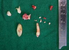
Notably, the CBCT indicated preservation of bony integrity around the lesion and ascertained that the surrounding neurovascular structures, notably the inferior alveolar nerve, were unaffected. Based on the radiographic and clinical examination, a compound odontome with an impacted left mandibular canine was diagnosed. The odontome was to be surgically enucleated along with the extraction of the affected tooth 32. The operation was performed under local anaesthesia with 2% lignocaine with 1:100,000 epinephrine. Povidone-iodine solution was used to disinfect the perioral region. A rectangular mucoperiosteal flap was raised from the right mandibular lateral incisor to the left first premolar to expose the bone. A bony window was made close to the apex of tooth 72 with a round tungsten carbide bur in a slow-speed handpiece with continuous irrigation with sterile normal saline to prevent thermal damage. After the removal of the cortical bone, several denticles were seen in the cavity. Seven denticles of differing sizes and shapes, along with the overlying follicular tissue, were removed with caution. Tooth 32, which was pathologically mobile and in close approximation to the lesion, was also removed. The cavity was irrigated well with normal saline to remove any remaining debris. The wound was closed with 3-0 Vicryl sutures after repositioning the mucoperiosteal flap. The post-operative course was smooth, and the patient was instructed on routine post-operative care, including pain management and oral hygiene maintenance. Histopathological examination of the resected tissue was consistent with the diagnosis of compound odontome. The case highlights the significance of early radiographic assessment in diagnosing odontogenic lesions with unusual presentations, as well as the rare occurrence of compound odontomes in the anterior mandible.


Cause of compound odontome development [14]. Still, the origin of this hyperactivity in such cases is yet unknown. One intriguing aspect of odontomas is their significant variety in denticle count. Although earlier reports noted the removal of 4 to 37 denticles, a recent remarkable instance reported that as many as 232 denticles were removed during enucleation [17]. Under local anaesthesia and extraction of tooth 32-related to the odontome, in this case-surgical enucleation was performed. To exclude additional mixed odontogenic neoplasms, such as ameloblastic fibro-odontoma or odontoameloblastoma [18], the sample was sent for histological investigation. Although benign in origin, odontomas can cause disturbances in the regular eruption of teeth. These could manifest as delayed eruption, impaction, over-retention of primary teeth, or malalignment caused by tooth displacement [19]. These consequences underscore the importance of early identification and treatment. The appropriate examination of odontomas has benefited much from radiographic imaging, more especially, from Cone Beam Computed Tomography (CBCT). High-resolution pictures of the lesion and its connection with surrounding anatomical structures in space offered by CBCT maximise diagnosis and surgical planning [20]. For odontomas, surgical excision is the preferred therapy. Smallsized lesions can be eliminated uneventfully, but surgeons should be careful not to damage surrounding structures, particularly in Areas with close anatomical packing. Early intervention ensures superior long-term results by reducing the risk of deformity and promoting the natural eruption of impacted permanent teeth. Regarding the present instance, a follow-up was indicated to evaluate and treat tooth 33, which had not erupted following surgery.
For patients with odontomas, the issue can escalate due to the pain or the mobility of the teeth. One such patient, a 27-year-old female, expressed the discomfort with her left mandibular anterior region. The pain, along with the discomfort, brought about a lot of discomfort and uncertainty concerns to the patient. The radiographic findings of several tooth structures also complicated her case as this would require other forms of imaging like a CT scan to obtain satisfactory clarity to the extent of the issue. Additionally, having a missing tooth and a missing canine caused a lot of self-consciousness and frustration to the patient as well.
When patients were told of having a compound odontoma, and the need to perform an enucleation procedure, it indeed would bring a lot of worries. Even though the procedure was done with the patient under local anesthesia, many of having abnormal growths caused worries such as the recovery procedure. Complications such as the eruption of the impacted 33 tooth would also provide a lot of worries especially with future dental visits. These reasons are understandable, particularly the positive and encompassing emotions. Since there were also worries of a potentially uncomfortable recovery, this might bring the patient to the endwhile worries about the enucleation procedure.
Nevertheless, the anticipated benign results and restoration of regular functionality and the patient's gratifications following the osteosarcoma excision and the scheduled oversight of the affected promontory offer optimism for a constructive subsequent encounter.
In essence, odontomas can create major clinical problems if left untreated, even if they are benign and usually asymptomatic. Their varied appearance emphasises the need to include them in differential diagnosis, particularly in younger people with unerupted or impacted teeth. A thorough approach, including clinical, radiological, and histological examinations, ensures appropriate treatment and a favourable prognosis.