Oral Sphere
Journal of Dental and Health Sciences
Journal of Dental and Health Sciences
Received: 2025-07-01
Accepted: 2025-09-12
Published: 2025-10-01
Pages: 233-238
Background: HIV and AIDS remain one of the leading causes of global mortality, with significant socio-economic and psychological implications. Oral manifestations are essential for diagnosing HIV, as early detection of these lesions can identify the progression to AIDS.
Case Presentation: A 45-year-old female patient presented with oral complaints, including gum pain, weight loss, fever, and skin lesions. Clinical examination revealed pseudomembranous candidiasis and linear gingival erythema. HIV diagnosis was confirmed through HIV antibody testing and CD4 count. The patient underwent a comprehensive clinical examination and relevant laboratory tests to assess both oral health and HIV status. HIV testing was performed using multiple assays, with a reactive result. A complete blood count (CBC) and enzyme-linked immunoassay (ELISA) test were conducted to identify HIV-related oral manifestations. The patient's CD4 count was reduced to 222 cells/mm³, indicating an impaired immune system. The observed oral lesions were consistent with HIV-related pseudomembranous candidiasis and gingivitis. After initiating Highly Active Antiretroviral Therapy (HAART), partial resolution of the lesions was observed within 20 days of treatment.
Conclusion: Oral lesions, especially candidiasis, are an early indicator of HIV infection. Dentists play a vital role in detecting HIV, which enables early intervention and timely treatment. Early recognition and treatment of oral manifestations are crucial in improving the prognosis of HIV-positive patients, particularly in resource-limited settings where advanced diagnostic tools may be unavailable.
HIV and AIDS have been accounted as the significant cause of mortality worldwide. AIDS and infection caused by HIV are one of life's most serious risks due socio-psycho-economic economic character [1]. Since 1981, several oral diseases related to AIDS caused by HIV have been since then. There are primarily two laboratory indicators i.e, CD4+ lymphocyte count and HIV viral load; however, any impoverished nations do not have access to these assays [2]. Therefore, clinical findings are used to screen the disease process. With the advent of new diagnostic tools clinicians and researchers have easier access to the or have access to the oral cavity, which is more straight forwarder [3]. Detection of oral lesions plays an essential role in the progression to AIDS as superinfections take over in an immunocompromised state [4]. According to various studies on this condition, oral lesions are observed in 70–90% of the patients who have HIV at any stage [5]. Most common lesions include oral candidiasis, linear gingival erythema, hairy leukoplakia, aphthous ulcer, kaposi sarcoma, and necrotising ulcerative periodontitis, along with a few less common ones [6]. Our article presents a case report of a female involving oral manifestations of HIV.
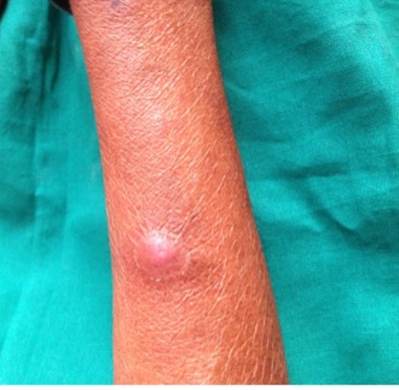
A female patient, 45 years old, came to us presenting pain in the upper and lower gingiva of her jaw. The medical history revealed skin allergy and itching on arms and legs, for which the patient has been taking medication for 1 month. The patient gave a history of a hysterectomy 2 years back due to pain in the pelvic region. The patient also gave a history of fever and weight loss of about 6-7 kgs in the last 2 months. The patient did not provide a history of a persistent cough or any other systemic diseases. The patient is married and has four children, with the youngest being 10 years old. The patient revealed the positive exposure of her husband, stating that her husband is a driver by occupation, has a habit of consuming alcohol daily, and has multiple sex partners. On general examination, a dome-shaped lesion was present on an arm, approximately 1 cm in diameter, and was erythematous, having a stretched and shiny surface with distinct features. It was soft and tender on palpation Figure 1.
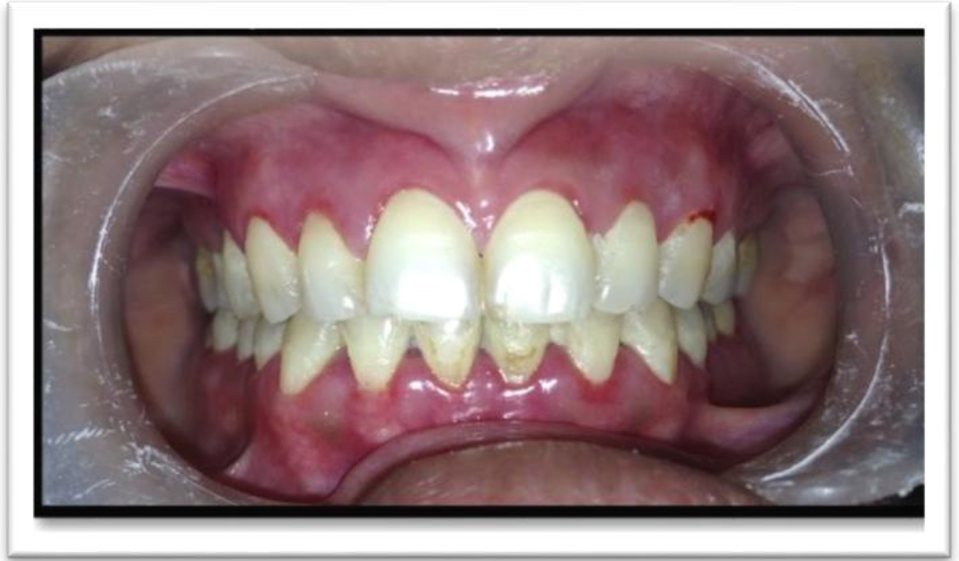
Also, multiple crops of cutaneous red itchy lesions were seen on legs of size approximately 0.5- 1 cm. The lesion appeared to be in the healing stage due to the presence of crusting. Temporomandibular joint movements were well coordinated and bilaterally symmetrical Bilateral submandibular lymph nodes, one in number, were palpable in size approximately 5 mm. Lymph nodes were firm in consistency, mobile and were tender. On intraoral examination inflammation of the marginal gingiva was seen exhibiting a distinct erythematous band of 2-3 mm in width with diffuse erythema extending to attached gingiva w.r.t maxillary anterior region Figure 2.
Also, plaque-like yellowish white coated appearance was seen on the tongue’s dorsal surface along a region of erythema of size approximately 30 mm having irregular margins in the anterior onethird of the tongue dispersed with a few ulcerations of size approximately 2×2mm. The surface appears to be smooth and glossy in the anterior one-third of the tongue and is devoid of filiform papilla. The lesion was partially scrapable having a smooth surface with irregular margins and was tender on palpation Figure 3.
Clinically, a final diagnosis of pseudomembranous candidiasis and acute marginal gingivitis was made. She was prescribed a topical application of Candid gum paint for 2 weeks, three times a day followed by a complete blood count (CBC) and enzyme-linked immunoassay (ELISA) test for HIV. The result of the HIV antibody test came out to be reactive Table 1. In treating the oral manifestations of HIV, several key objectives must be pursued to ensure comprehensive care and optimal outcomes following diagnosis. A routine blood investigation revealed CD4 count reduced to 222 cells/mm3, whereas the normal value is 500-1200 cells/mm3, and the HIV kit tests gave a positive result. These investigations lead to a final diagnosis of HIV-induced pseudomembranous candidiasis. The patient was referred to the Antiretroviral Therapy (ART) Department of Safdarjung Hospital, New Delhi. A complete medical workup was done, and Highly Active Antiretroviral Therapy (HAART) was initiated, which included cotrimoxazole, zidovudine (300mg) + famciclovir (150mg) + Nevirapine (200mg) twice daily for an initial period of 15 days. The medication dosage was individualised to the patient's requirements. On follow-up after 20 days of HAART therapy, the lesion had partially resolved.


| Date: | 4/5/2025 | |
| Age/Sex: | 45/F | |
| Pid No. : | SGT 510 | |
| Date when sample was received: | 4/5/2015 | |
| Result of HIV antibody test: | Reactive | |
| First test (Name of the kit) | Alere Trueline | HIV-1/HIV-1&2 Reactive/ Non-reactive |
| Second test (Name of the kit) | SD Bioline | HIV-1/HIV-1&2 Reactive/ Non-reactive |
| Third test (Name of the kit) | Tridot | HIV-1/HIV-1&2 Reactive/ Non-reactive |
HIV infection presents varied oral manifestations shown by patients at some point of their disease. As discussed above the most common of all oral manifestations is candidiasis. In our case, the patient reported in a relatively healthy state and was completely unaware of her immune status. The patient presented with the typical features of pseudomembranous candidiasis including a burning sensation along with acute marginal gingivitis, and these findings triggered investigations for HIV infection. Mostly the diagnosis of HIV is made after discovery of these oral lesions, especially candidiasis.
Infections in HIV are caused due to the immunosuppressive state brought about by reduced absolute CD4 levels and a deranged cytokine network, both of which have devastating clinical effects. HIV infection has a wide range of clinical effects, from an acute sickness linked to the original infection to a protracted asymptomatic condition to advanced disease. When any HIV patient presents, their dental condition is a crucial indicator of their overall health since it may provide valuable insights into their immune system [7]. These conditions can manifest as a variety of lesions, including bacterial, viral, and fungal infections; malignant neoplasms like Kaposi's sarcoma; and nonspecific presentations including aphthous ulcerations and salivary gland disease. Factors such as CD4 counts less than
200 cells/mm3, viral load greater than 3000 copies/mL, dry mouth/xerostomia, poor oral hygiene, and smoking or perhaps any habit as such predisposes expression of oral lesions [8].
Oral candidiasis is the most common illness associated with HIV infection; it impacts 17–43% of HIV-positive individuals and more than 90% of AIDS cases. Most people with significant untreated HIV infection typically suffer from oropharyngeal candidiasis, which is one of the first signs of HIV-induced immunodeficiency. As it develops earlier than other serious opportunistic infections, it could be a significant indicator on the onset or advancement of infections associated with HIV [9].
There are four primary kinds of Candidiasis infection namely hyperplastic candidiasis, erythematous candidiasis, pseudomembranous candidiasis, and angular cheilitis. Subjects usually display any one of these appearances, or less commonly a mix of them. While pseudomembranous form of candidiasis being more prevalent in people with AIDS, the erythematous form is more common in those infected with HIV [10]. Red, macular lesions are the classic presentation of erythematous candidiasis, usually seen on the tongues’s dorsum and hard palate.
The hyperplastic type is characterized by white plaques common in the buccal mucosa and cannot be removed by scraping. The representation of pseudomembranous candidiasis appears as ‘creamy white curd-like plaques’ on oral mucosal surfaces that can be wiped away, leaving a red or bleeding underlying surface. Cases of angular cheilitis present as ulcerations, peeling or cracking inside the commissure of the lip or corner of the mouth also frequently present in combination with other forms of candidiasis [11].
Rare infections like histoplasmosis can also aid diagnosis of the condition. Tuberculosis, another prominent bacterial infection, seen as a coinfection in AIDS, commonly in the Indian population where tuberculosis is the most common air borne infection [12].
Candidiasis can be diagnosed by detection of fungal pseudohyphae in exfoliative cytologic preparations, frequently using Schiff and/or Papanicolaou-stained (PAP Stain) preparations on a periodic basis. However, pseudomembranous candidiasis has the highest yield of positive cytology smears [13].
A lower CD4:CD8 ratio and an increase in the severity of HIV infection are associated with a higher frequency of candida species isolation. CD4 cell count < 200 cells/mm3 is strongly linked with oral symptoms, particularly candidiasis. In case of mild to severe infections, topical treatments including nystatin oral suspension and pastilles or clotrimazole troches can be used. Fluconazole, the most often used medication. Other antifungals like itraconazole, and voriconazole are a couple of systemic medicines that are saved for cases of moderate to severe illness for fluconazole-resistant patients. Patients with HIV tend to develop both oral and esophageal candidiasis, necessitating a longer course of treatment with larger dosages of antifungals [14].
Unquestionably, the patient's angular cheilitis, periodontitis, and erythematous candidiasis, together with his lack of response to topical antifungals, were the main reasons that probed us about his lifestyle and conducted the tests that ultimately resulted in an HIV diagnosis.
Oral lesions as discussed serve as an early label for HIV infection and may indicate decline in general health and a poor prognosis. The oral lesions are a reliable indicator of HIV infection and might indicate a decline in overall health and a dismal outlook. In order to enable early HIV detection and ensure prompt medication start, the dentist must possess a thorough understanding of the traits and presentation of HIV infection symptoms. The initial clinical indication of HIV infection might frequently be a candidal infection. If an otherwise healthy
person develops oral candidiasis, lifestyle and other factors related to HIV infection should be investigated. Dentists play an important role in detecting presentation of the manifestations of HIV infection, which aids early identification of HIV, and making early intervention possible. Therefore, in underdeveloped nations like India, with one of the highest prevalence of HIV infection where RNA load assessment and CD4 count are usually unfeasible due to high costs in large populations, oral symptoms can serve as a measure of immune state. As a result, oral lesions linked to HIV are thought of as "sentinels and signposts" of HIV/AIDS, as in our case as well. Early detection and treatment of these lesions are crucial to preserving the health and extending the lives of AIDS patients.
To my surprise, at 45 years old and female, I never thought I would receive a diagnosis of HIV. I had come to the clinic because I had concerns regarding oral issues, including, gum pain, pain, and skin lesions, along with fever and weight loss. I was surprised to learn that such a constellation of symptoms could be indicative of HIV. I crossed the HIV antibody test. Along with a decrease in my CD4 count and the diagnosis of pseudomembranous candidiasis with acute gingivitis, I had my answer. Although the diagnosis was overwhelming, I was able to find a sense of relief in the fact that my condition was one that could be managed with the help of HAART (Highly Active Antiretroviral Therapy) because I was still in the early stages of the disease. Over the next several weeks, I was able to start regaining hope as some of my symptoms began to improve. I really appreciated the early role that dentists played in my diagnosis. I also learned that I should seek help as soon as I am faced with some unusual oral issues. I appreciate all the support and the treatment plan that I was able to receive.