Oral Sphere
Journal of Dental and Health Sciences
Journal of Dental and Health Sciences
Received: 2025-10-29
Accepted: 2026-01-30
Published: 2026-04-01
Pages: 110-114
Background: Mulberry molars are typically representing oral signs of congenital syphilis, characterized by multiple globular enamel cusps on the occlusal surfaces of first permanent molars. But sometimes, these morphological features can be seen in persons who never had syphilis or physiological evidence of it.
Case Presentation: A case of a 12-year-old female patient presenting with characteristic mulberry molars involving all four first permanent molars. The patient’s medical and family history was non-contributory, and both Venereal Disease Research Laboratory (VDRL) and Treponema pallidum hemagglutination tests were negative, ruling out congenital syphilis. On clinical examination, multiple globular cuspal protuberance without caries or occlusal interference. Conservative management was followed, emphasizing periodic monitoring and topical fluoride application. This case highlights the possibility of non-syphilitic etiologies including local, systemic, genetic, or environmental condition which is a contributing factor for enamel developmental defects resembling mulberry molars.
Conclusion: Mulberry molars can occur irrespective of congenital syphilis. It is important to know this rare occurrence for diagnostic purposes. Thus, this particular case requires teamwork between the dentist and the paediatrician to avoid diagnostic errors.
Enamel developmental defects represent a heterogeneous group of anomalies involving the size, shape, thickness, and mineralization of the tooth enamel, and often functional and aesthetic concerns [1]. Among these defects, mulberry molars are a distinctive morphological abnormality characterized by multiple globular enamel cusps replacing the normal occlusal anatomy of first permanent molars [2]. Classically, mulberry molars are regarded as pathognomonic dental stigmata of congenital syphilis, a vertically transmitted systemic infection caused by Treponema pallidum that affects enamel formation during early tooth development [3].
The enamel hypoplasia that produces the characteristic “mulberry” appearance includes irregular, rudimentary enamel cusps with deficient, disorganized enamel, often accompanied by diminished tooth size and altered occlusal morphology [4]. Typically, such defects are identified during late childhood when the first permanent molars erupt and are associated with a history of maternal syphilis or positive serological markers [5].
However, a few recent reports question the occurrence of congenital syphilis as the etiological factor for mulberry molars. Few cases with similar enamel characteristics have also been reported. developmental defects in the form of mulberry molars, in patients without clinical, serological, or historic evidence of syphilis [6],[7].
This case remains relevant in contemporary dental literature because world challenges still exist in distinguishing syphilitic lesions of the enamel from non-syphilitic lesions in the increasing rates (nearly 4,000 US cases in 2024 - up 700% since 2015), emphasizing the importance of comprehensive diagnostic methods other than historical linkage [8].
Distinguishing between true syphilitic mulberry molars from non-syphilitic enamel anomalies is important for accurate diagnosis, prognosis, and management. This rare case of mulberry molars in a 12-year-old female with negative serological tests and no maternal history of syphilis is presented to emphasize the possibility of non-syphilitic causes and to stress the role of comprehensive clinical and laboratory evaluation.
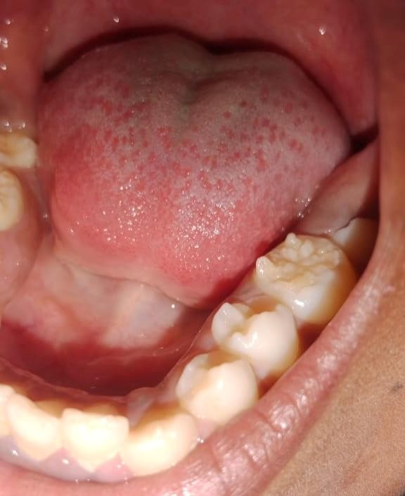
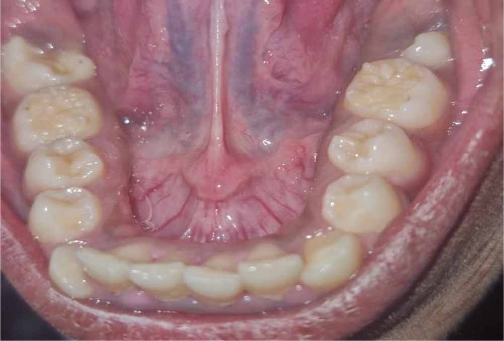
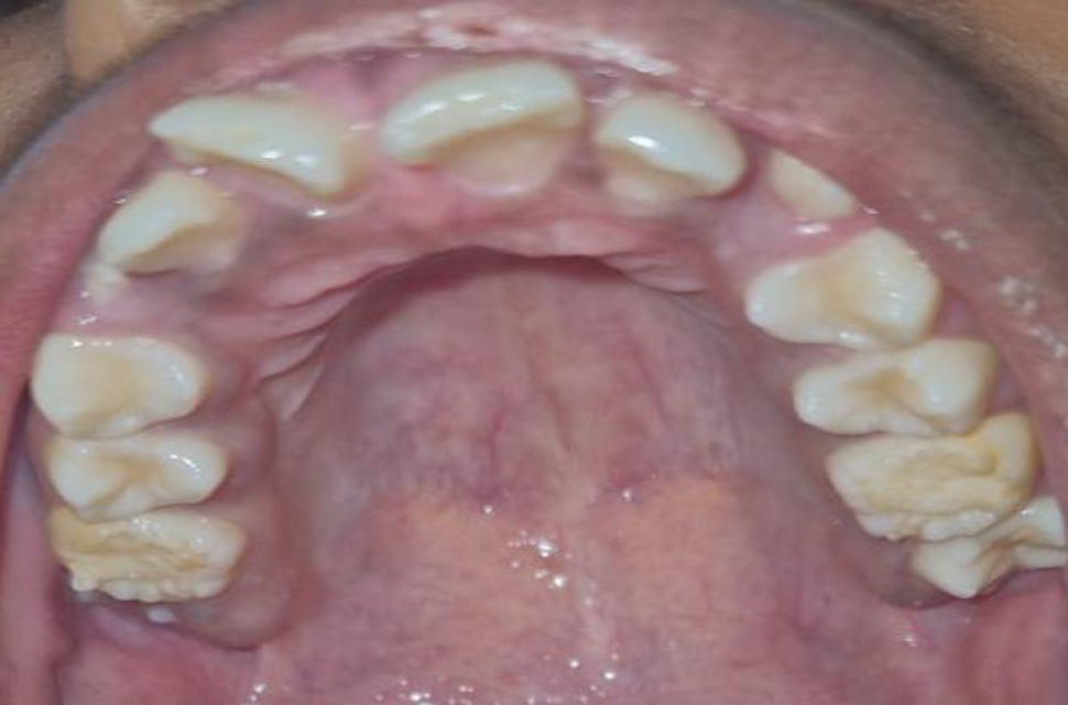
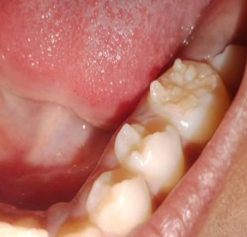
In a 12-year-old female patient an uncommon enamel defect characterized by multiple globular-shaped cusps on the occlusal surfaces of all four first permanent molars, results in a typical "mulberry" appearance Figure 1 and Figure 2.
This morphological anomaly resembled mulberry molars, which are classically seen in congenital syphilis - a condition transmitted from mother to child during pregnancy causing enamel hypoplasia and stunted molars with disorganized enamel cusps. However, there were no maternal history of syphilis or fever during pregnancy. Serological tests such as Venereal Disease Research Laboratory (VDRL) test and Treponema pallidum hemagglutination assay (TPHA) were both negative, effectively ruling out congenital syphilis.
The past medical and family history was unremarkable, with no similar dental abnormalities in parents / siblings, and no signs of systemic illness.
Clinical examination showed the presence of multiple globular-shaped cusps on the occlusal surfaces of the teeth. The upper and lower first permanent molars, with normal Class I molar relationship Figure 3. These characteristic cuspal projections were rounded, did not interfere with occlusion, caused no discomfort. The molars were free of caries and functional.
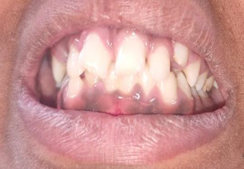
Follow-up and Management: The patient was monitored over a 24-month follow-up period (from initial presentation through regular 6-monthly reviews), during which the teeth remained functional, asymptomatic, with no progression of enamel defects, and no caries development. Conservative management included topical fluoride application to strengthen enamel and periodic clinical monitoring. The patient's overall dental and craniofacial growth/development progressed normally for chronological age, with no deviation from expected norms Figure 4.
Mulberry molars are classically recognized as pathognomonic dental stigmata of congenital syphilis, characterized by multiple small, rounded enamel cusps creating a "mulberry-like" occlusal surface on first permanent molars due to enamel hypoplasia from Treponema pallidum infection [9]. In late congenital syphilis, these dwarfed molars form alongside Hutchinson's incisors and interstitial keratitis [10].
However, recent literature challenges this exclusive association, documenting non-syphilitic mulberry molars classified as "developmental enamel defects with mulberry morphology" or "non-syphilitic enamel hypoplasia variants" [11]. In our case, all four first permanent molars exhibited classical mulberry morphology without serological evidence of syphilis, normal occlusion, and no associated stigmata.
Amelogenesis imperfecta (AI) was ruled out due to: (1) localized distribution (only first permanent molars vs. AI's generalized enamel involvement), (2) normal enamel thickness elsewhere, (3) absence of familial pattern, and (4) normal radiographic dentin/pulp morphology (AI typically shows thin enamel with contrasting dentin). Turner's hypoplasia excluded due to bilateral symmetrical involvement of all first molars (Turner's = single tooth/asymmetrical from primary tooth trauma) [13],[14] Table 1 shows the comparison with published Non-Syphilitic Cases Table 1.
| Case | Year | Age | Teeth Affected | Serology | Occlusion | Follow-up |
|---|---|---|---|---|---|---|
| Koneru et al [4] | 2019 | 6y/ M | All 4 first permanent molars | Negative VDRL/TPHA | Normal | Not reported |
| Harizanova et al [5] | 2024 | 18yrs/F | First mandibular Second permanent molar | Negative | Normal | Periodic monitoring |
| Darwin & Castelino [11] | 2023 | 17y M | Multiple permanent molar | Negative | Normal | Not reported |
| Present case | 2025 | 12y F | All 4 first PMs | Negative VDRL/TPHA | Class I | 24 months |
The Etiological Considerations of Non-syphilitic mulberry molars likely arise from multifactorial enamel hypoplasia during the late bell stage of odontogenesis (8-12 months’ gestation):
Local: Primary tooth germ infection/trauma
Systemic: Nutritional deficiency, hyperthermia, hypoxia
Genetic: Subtle amelogenesis gene variants
Environmental: C-section delivery (altered ameloblast oxygenation) [12],[13].
The Current Classification of Non-syphilitic cases fall under "localized developmental enamel defects" per Pindborg classification, distinct from generalized Amelogenesis imperfecta [14],[15].
As a patient, my experience at the dental clinic was primarily for a routine dental check-up. I was not experiencing any pain, eating difficulties, or any other dental problems. During the check-up, the dentist observed that the biting surfaces of my molar teeth appeared different from normal teeth. Prior to this, I was not aware of any unusual features about my teeth since they were functioning properly.
The dentists clarified that my teeth appeared as if they had a condition that is normally associated with some infections. This piece of information initially alarmed me and my parents.
However, the dentists took time to clarify the need for blood tests to establish a diagnosis and that this was only a means to exclude potential causes. Once the results showed that I was normal, we were relieved and comforted.
The dentists clarified that since my teeth were healthy, with no decay and not causing any problem with biting or chewing, there was no need for extensive treatment. A conservative treatment method was recommended. This involved the use of topical fluoride to make the enamel stronger and regular check-ups every six months to monitor the teeth.
During the follow-up visits, the doctors closely examined my teeth and made sure that there were no changes, pain, or cavities. Over time, my teeth remained comfortable and fully functional, which made me feel confident and safe with the treatment plan.
This experience has made me realize the importance of visiting a dentist regularly, even if there are no symptoms. I am thankful to the dental team for their careful evaluation, explanations, and approach that reduced my anxiety and ensured that I received proper care without any unnecessary treatment.
Non-syphilitic mulberry molars are a kind developmental enamel anomalies requiring comprehensive clinical and serological assessment beyond traditional syphilis association. This case represents an excellent 24-month prognosis with conservative fluoride treatment and periodic monitoring, establishing clear management criteria for functional, asymptomatic presentations.
Interdisciplinary involvement of pediatricians for maternal/systemic history, pediatric dentists for clinical assessment and oral pathologists for confirmatory evaluation when indicated will help to avoid misdiagnosis, social stigma, and ensures holistic patient care amid rising congenital syphilis prevalence. This report has wider clinical implications as it establishes the stability of non-syphilitic cases over a period of time, thus facilitating the confirmation of the conservation protocols used. Future studies should be directed towards etiology as well as outcomes of stability of the occlusal plane.