Oral Sphere
Journal of Dental and Health Sciences

Journal of Dental and Health Sciences
Received: 2026-05-12
Accepted: 2026-08-02
Published: 2026-10-01
Pages: 263-267
Background: A nasopharyngeal hamartoma (NH) is a very rare congenital, benign condition in which there is an unorganized growth of mature tissues found at the site of the lesion. Hamartomas are typically found in organs like the liver, lungs and kidneys, but when the adult nasopharynx is involved, it is very rare and diagnosis and management are important.
Case Presentation: A 42-year-old man with occasional nasal congestion was incidentally found to have a 10-mm nasopharyngeal polyp during routine endoscopy. The computed tomography, magnetic resonance imaging and narrow-band imaging showed a well circumscribed lesion with no malignant features. It was totally resected endoscopically with general anesthesia. Histopathological examination showed pseudostratified columnar epithelium, with a small amount of salivary gland tissue, adipose tissue and smooth muscle fibers, which confirmed the diagnosis of nasopharyngeal hamartoma. Post-operative course was uneventful.
Conclusion: Adult NH is a very unusual condition that can be found by chance and have mild symptoms. Full endoscopic, radiological and histopathological examination is important for proper diagnosis and exclusion of malignancy. Complete endoscopy is an effective treatment and prognosis is excellent, with very low risk of recurrence.
Hamartoma is a developmental anomaly in which mature tissue elements of the organ where it forms grow in a disorganized fashion. Hamartomas are differentiated cells with normal cytological features, but an abnormal architectural pattern is seen, unlike true neoplasms [1]. They are regarded as examples of malformations which are not acquired tumors, but rather examples of the abnormal development of the embryo. Hamartomas have limited growth potential but can gradually increase in size and become clinically important depending on the size and location of the hamartoma [2]. They are often benign with slow progression and the diagnosis can be difficult, especially if the tumors are located in unusual places or have vague symptoms [3].
Lung, liver, spleen, kidneys, pancreas and hypothalamus are the common sites of hamartoma occurrence, and the most common is incidental detection by imaging or histopathology procedures for different reasons [4]. Hamartomas of the head and neck area, however, are rare and make up a minority of cases reported. In the upper aerodigestive tract, these lesions are known to occur in the nasal cavity, paranasal sinuses, oral cavity, larynx, and nasopharynx, and the latter is one of the least common sites of occurrence [5]. As a result of their rarity, especially in adults, clinicians might not consider hamartoma as a differential diagnosis in the diagnosis of nasopharyngeal masses initially [6].
Presumably, nasopharyngeal hamartomas occur mostly in infancy and childhood, when symptoms of upper airway obstruction such as nasal blockage, feeding problems, recurrent upper respiratory tract infections, snoring, or sleep disordered breathing are found [7]. There are only small case series and isolated case reports of adult cases in the literature and they are very rare. In adults these lesions are often non-specific such as intermittent nasal congestion, a sense of a foreign body, post-nasal drip, and headache, and may only be discovered on routine endoscopy or are incidental findings [8]. Due to their age-specific nature and sensitivity to clinical signs, adult nasopharyngeal hamartomas may be confused with inflammatory polyps, epipharynx cysts (Tornwaldt cysts), respiratory epithelial adenomatoid hamartoma (REAH), benign mesenchymal tumors or even with malignant lesions such as nasopharyngeal carcinoma and nasopharyngeal lymphoma [9].
Radiological assessment by computed tomography (CT) and magnetic resonance imaging (MRI) allows detailed information about the size, extent, anatomical composition, and adjacent structure involvement of the lesions to be obtained. However, imaging findings are frequently not enough to make a clear diagnosis as there is significant overlap between benign and malignant nasopharyngeal lesions [10]. Therefore, the histopathological examination is the gold standard diagnosis and shows a disorganized mixture of mature tissue elements such as adipose tissue, minor salivary glands, smooth muscle, fibrous tissue, and respiratory epithelium that is devoid of cytological atypia and invasive growth [11]. Complete surgical removal, ideally by an endoscopic transnasal technique, is recommended and has very small morbidity, very low recurrence rates and excellent long-term results [12]. This report presents a rare case of nasopharyngeal hamartoma diagnosed by thorough clinical, radiological and histopathological work-up and emphasizes the need to include this rare but benign entity in the differential diagnosis of nasopharyngeal masses and to reappraise the current evidence on the diagnosis and management of such lesions.
A 42-year-old male underwent a routine health assessment, during which an upper gastrointestinal nasal endoscopy identified a nasopharyngeal polyp. He did not find it particularly troubling, but occasionally experienced nasal congestion. His medical history was unremarkable, with no notable familial or personal health issues, and he has no history of smoking.
Endoscopic evaluation of the nasopharynx revealed a smooth-surfaced, polypoid lesion approximately 10 mm in diameter, situated centrally on the posterior wall, slightly deviated to the left. Narrow Band Imaging (NBI) did not demonstrate any abnormal microvascular patterns or features suggestive of malignancy on the mucosal surface Figure 1.

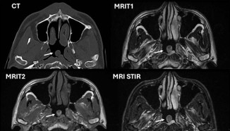
Computed tomography (CT) scans depicted a polypoid lesion measuring approximately 10 mm in diameter with a base width of 0.5 cm, located on the posterior wall of the nasopharynx, slightly to the left. The lesion exhibited an almost homogeneous internal structure Figure 2.

Magnetic Resonance Imaging (MRI) further characterized the lesion: T1-weighted images showed an irregular shadow with mixed intermediate to high signals internally, while T2-weighted images revealed a spherical lesion with a stalk, displaying mixed low to intermediate signals. The MRI Short T1 inversion recovery (STIR), fat suppression sequences, indicated slightly elevated signals, consistent with the lesion's composition.
Under general anesthesia, the lesion was visualized using a rigid nasal endoscope. Submucosal injection of 1% xylocaine with epinephrine was administered to facilitate dissection. The stalk of the polyp was carefully separated using electrocoagulation, resulting in minimal intraoperative bleeding.
The excised tissue was identified as a polypoid lesion covered with pseudostratified columnar epithelium. Histological examination revealed the presence of minor salivary glands, adipose tissue, and smooth muscle fibers, leading to a diagnosis of hamartoma, a benign, disorganized proliferation of tissue native to the site Figure 3.

Hamartomas are benign, tumor-like malformations characterized by the disorganized proliferation of tissue components that are normally present within an organ. First described by Albrecht in 1904, these lesions are typically congenital anomalies resulting from abnormal tissue development during embryogenesis [13]. Although hamartomas are most frequently observed in organs such as the lungs, spleen, liver, kidneys, and pancreas, their occurrence in the head and neck region, particularly within the nasopharynx, is notably rare [14].
Patients with nasopharyngeal hamartomas may present with nonspecific symptoms such as airway obstruction, cough, headaches, and postnasal drip. Notably, some lesions are asymptomatic and are discovered incidentally during routine examinations. Endoscopic evaluation typically reveals a soft, submucosal mass with a smooth surface, often mistaken for inflammatory polyps [15]. Imaging modalities such as computed tomography (CT) and magnetic resonance imaging (MRI) provide further insights: CT scans may show partial calcification within the lesion, while MRI often reveals an irregular mass with heterogeneous signal intensities, including partially high on T1-weighted images and areas of low signal on T2-weighted images [16].
Differentiating nasopharyngeal hamartomas from other neoplastic and benign lesions requires comprehensive imaging and endoscopic assessment. Differential diagnoses include Tornwaldt cysts, lipomas, vascular fibromas, teratomas, nasopharyngeal carcinoma, and malignant lymphomas. Due to overlapping features, definitive diagnosis often necessitates histopathological examination, which reveals tissue components such as adipose tissue, salivary gland tissue, and smooth muscle. The presence of salivary gland structures within the lesion suggests heterotopic tissue proliferation rather than true neoplasia [17].
Hamartomas, which refer to the presence of normal tissue in an abnormal location, are classified into epithelial, mesenchymal, or mixed types. The epithelial adenomatous hamartoma is the most prevalent subtype in the nasal cavity and nasopharynx. The hamartomas present as respiratory epithelial adenomatoid hamartomas (REAH) in the nasopharyngeal region [18]. These lesions are characterized by the proliferation of respiratory epithelium forming nodular structures, which frequently have a smooth surface resembling inflammatory polyp. Mesenchymal hamartoma, although rarer, includes subtypes such as chondroid, chondromatous, vascular, lipomatous, and smooth muscle hamartoma. Histologically, the hamartoma presenting a significant component of smooth muscle tissue is infrequent. Literature reviews indicate that smooth muscle hamartomas in the nasopharynx are exceedingly rare, with only a single documented case reported to date [19]. The subtype of our case was diagnosed as smooth muscle hamartoma, likely resulting from heterotopic proliferation of smooth muscle tissue during early development.
Current treatment strategies primarily involve surgical excision, with transnasal approaches increasingly favored due to their minimally invasive nature. The prognosis following complete removal is excellent, with no reported cases of recurrence or malignant transformation associated with heterotopic lesions in the head and neck region, aside from REAH [20]. The pathogenesis of REAH remains incompletely understood. Nonetheless, surgical treatment combined with diligent follow-up is recommended to monitor for potential recurrence or complications, ensuring optimal patient outcomes [21].
I had no significant symptoms except occasional nasal congestion and was surprised when a routine health check revealed a small growth in my nasopharynx. Although I was initially anxious about the possibility of cancer, the medical team explained the findings clearly and performed further investigations before recommending endoscopic removal. The surgery was smooth, my recovery was uneventful, and I was relieved to learn that the lesion was a benign hamartoma. I am grateful for the early diagnosis and treatment, and I appreciate the thorough care provided by the multidisciplinary team, which gave me confidence and reassurance throughout the process.
This case exemplifies a nasopharyngeal hamartoma presenting as a polypoid lesion, diagnosed through comprehensive endoscopic, radiologic, and histopathological evaluation. The management involved minimally invasive surgical excision with favorable intraoperative findings and histological confirmation, underscoring the importance of multidisciplinary assessment in sinonasal lesions.